


Lightweight Medical Image Segmentation Incorporating Gaussian Mapping and Adaptive Feedforward
-
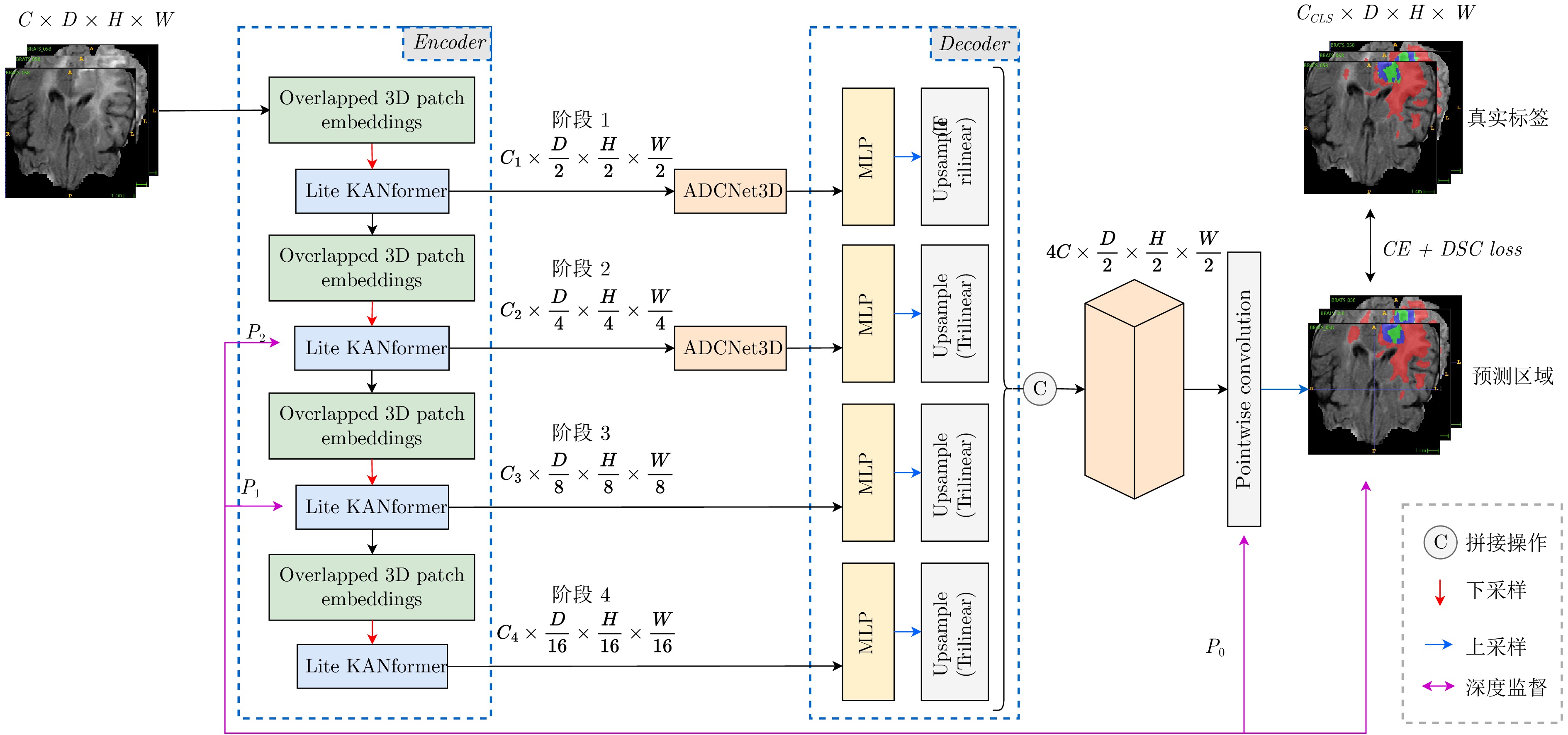
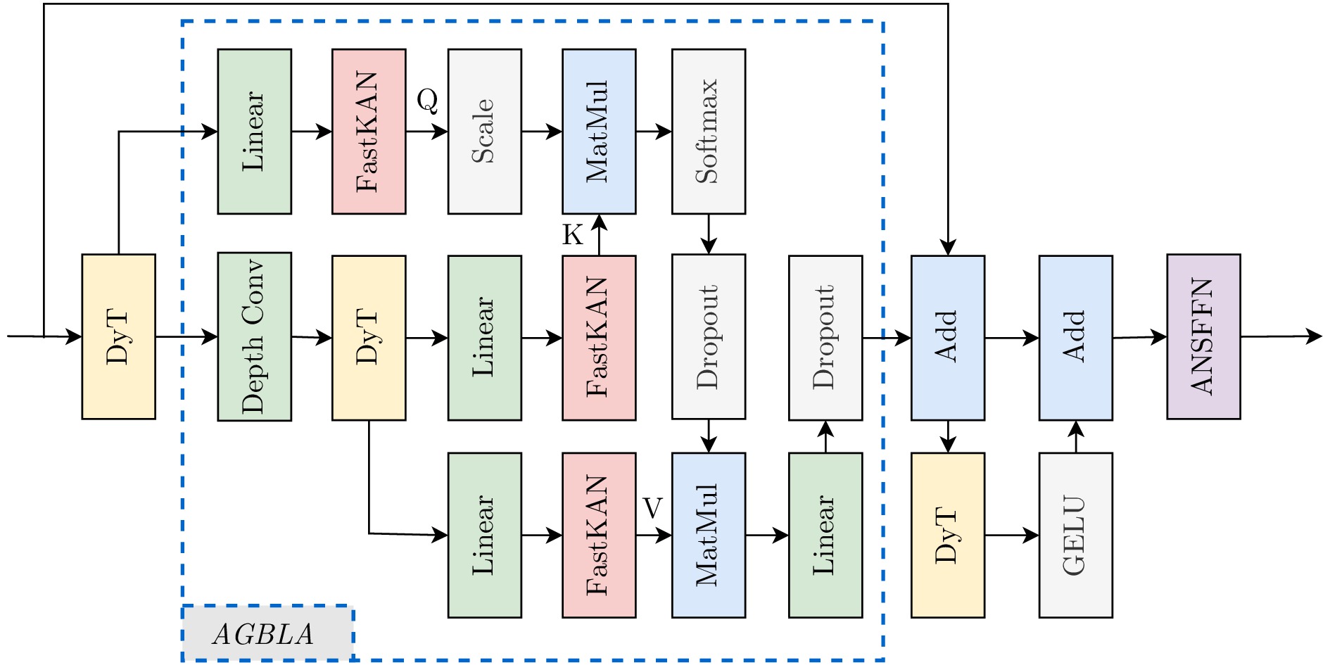
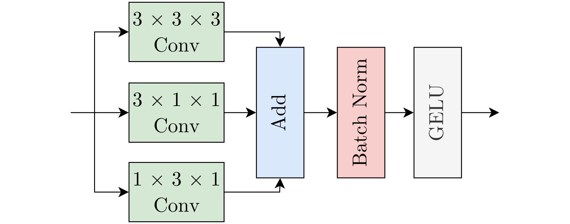
摘要: 在医学图像分割领域, 传统基于卷积神经网络(convolutional neural networks, CNN)的方法受限于局部感受野, 难以捕获全局信息, 而基于视觉转换器(Vision Transformer, ViT)的方法虽能通过自注意力机制捕获全局信息, 但需对图像Patch化处理, 导致模型难以捕捉跨块和块内的边缘纹理细节, 因此如何实现两者间的优势互补, 平衡精度与效率尤为重要. 为解决该问题, 提出一种融合高斯映射和自适应前馈的轻量医学图像分割方法—LiteSegFormer3D. 在编码器部分采用融合高斯映射和自适应前馈的Transformer块, 并在浅层跳跃连接处采用非对称卷积, 二者协同扩展了模型的多尺度感知能力, 弥补了分割过程的局部信息损失, 解码器则沿用基线的全MLP架构, 规避了传统3D解码器高昂的计算开销. 在BraTS 2017、ACDC和Synapse三个公开经典数据集上与多种主流的CNN、Transformer及混合方法进行对比实验, 结果显示, 该方法在BraTS 2017上DSC提升3.5%, HD95降低0.65 mm; 在ACDC上DSC提升1.07%; 在Synapse上DSC提升2.76%, HD95降低10.59 mm. 尤为重要的是, 本方法的可训练参数仅为1.73M, 相较基线SegFormer3D缩减了61.64%. 该方法展现出优异的精度与性能平衡, 具备临床轻量化部署潜力. 代码:
https://github.com/Strivy-ZSY/litesegformer3d Abstract: In the field of medical image segmentation, traditional methods based on convolutional neural networks(CNNs) are constrained by their local receptive fields, making it difficult to capture global information. Meanwhile, methods based on Vision Transformers(ViTs) can capture global information through self-attention mechanisms, but their patch-based image processing makes it challenging for the model to capture edge texture details both across patches and within patches. Therefore, achieving complementary advantages between the two approaches while balancing accuracy and efficiency is crucial. To address this challenge, we propose LiteSegFormer3D—a lightweight medical image segmentation method integrating Gaussian mapping and adaptive feedforward. The encoder employs Transformer blocks integrating Gaussian mapping and adaptive feedforward, while shallow skip connections utilize asymmetric con-volutions. This dual approach synergistically enhances the model's multiscale perception capabilities, mitigating local information loss during segmentation. The decoder adopts the baseline's full MLP architecture, thereby avoiding the high computational overhead associated with traditional 3D decoders. Comparative experiments against multiple mainstream CNN, Transformer, and hybrid methods on three public benchmark datasets—BraTS 2017, ACDC, and Synapse—revealed that our approach achieved a 3.5% increase in DSC and a 0.65 mm reduction in HD95 on BraTS 2017; on ACDC, DSC improved by 1.07%; on Synapse, DSC increased by 2.76% while HD95 decreased by 10.59 mm. Crucially, the trained parameters of this method total only 1.73 million, representing a 61.64% reduction compared to the baseline SegFormer3D. This approach demonstrates an excellent balance between accuracy and performance, show-casing potential for lightweight clinical deployment. -
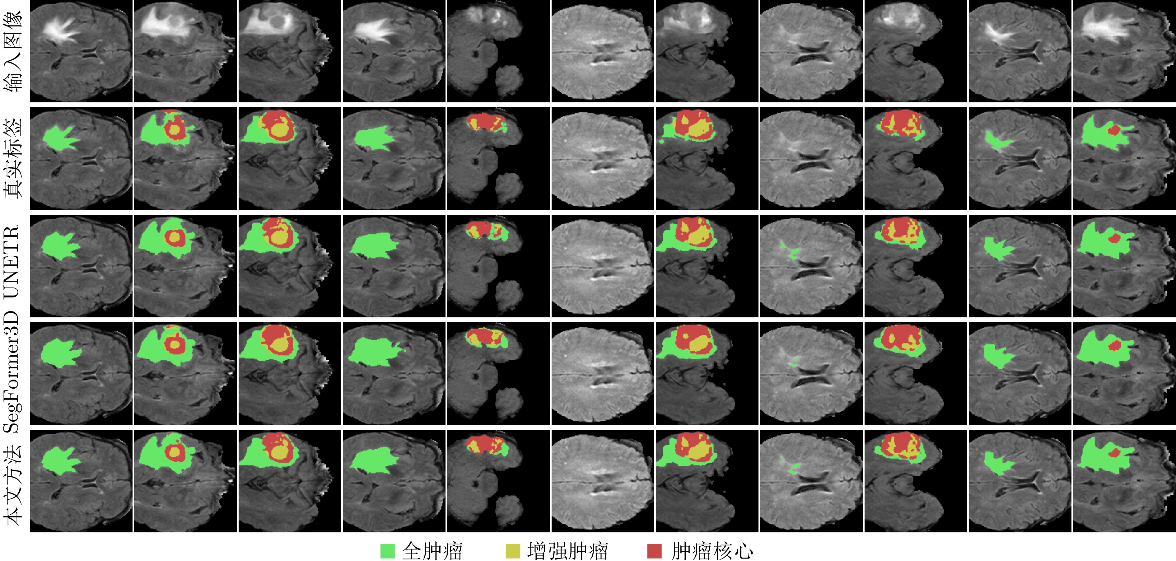
图 6 BraTS 2017数据集分割的可视化
注: 图中切片均取自同一例体数据, 为全面展示不同深度下的肿瘤形态变化, 切片采取随机抽样的方式.
Fig. 6 Visualization of BraTS 2017 dataset segmentation
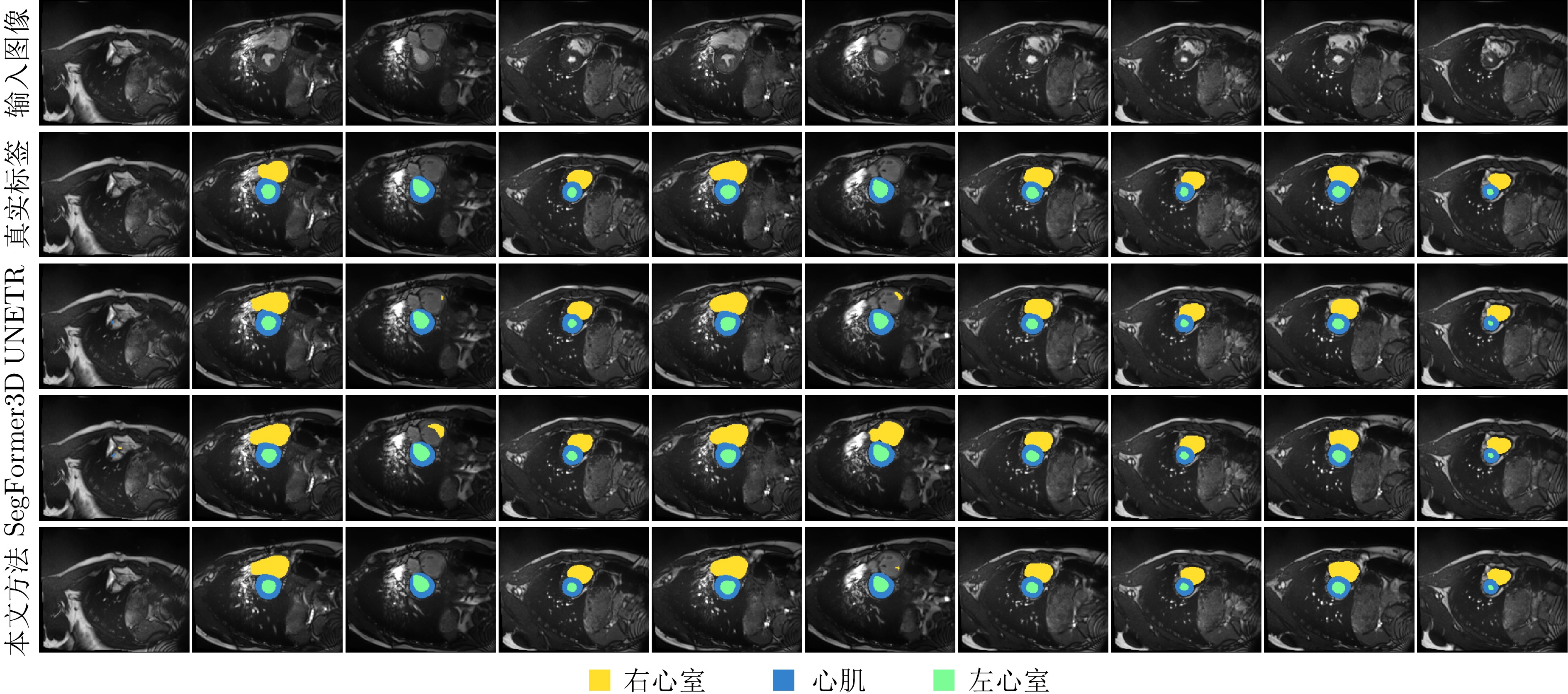
图 7 ACDC数据集分割的可视化
注: 图中切片均取自同一例体数据, 为全面展示不同深度下的器官形态变化, 切片采取随机抽样的方式.
Fig. 7 Visualization of ACDC dataset segmentation
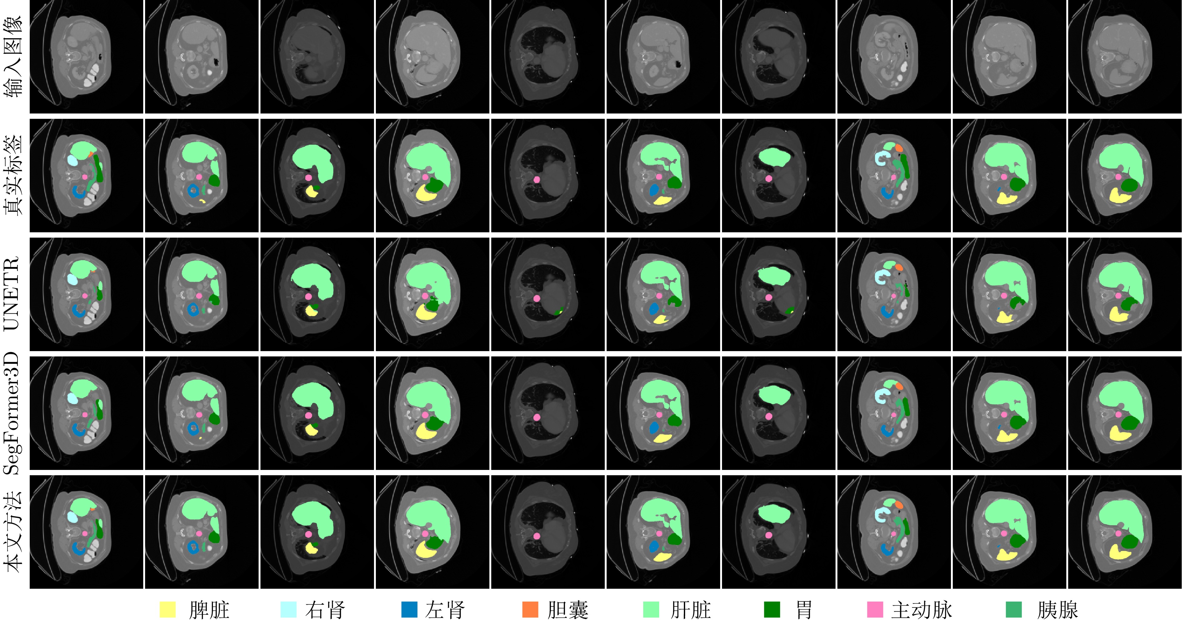
图 8 Synapse数据集分割的可视化
注: 图中切片均取自同一例体数据, 为全面展示不同深度下的器官形态变化, 切片采取随机抽样的方式.
Fig. 8 Visualization of Synapse dataset segmentation
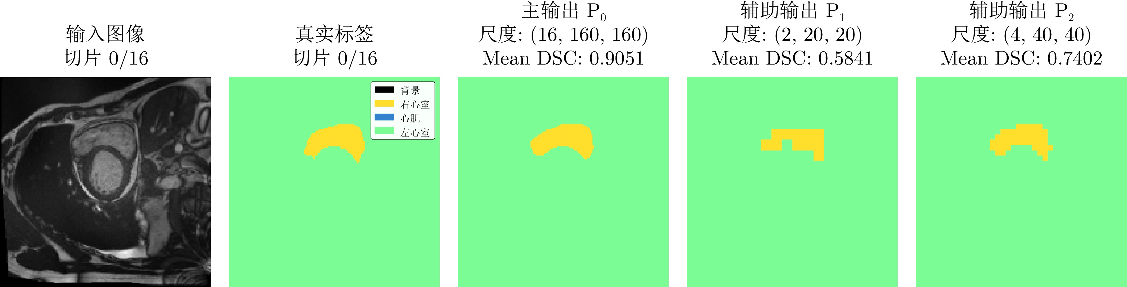
图 9 金字塔式深度监督过程可视化
注: 图中切片均取自同一例体数据, 为全面展示不同深度下的器官形态变化, 切片采取随机抽样的方式.
Fig. 9 Visualization of the pyramidal deep supervision process
表 2 在BraTS 2017数据集上与其他方法对比
Table 2 Comparison with other methods on the BraTS 2017 dataset
方法 年份 TP/M $ \downarrow $ 平均$ \uparrow $ 全肿瘤$ \uparrow $ 增强肿瘤$ \uparrow $ 肿瘤核心$ \uparrow $ DSC $ \uparrow $ HD95 $ \downarrow $ DSC $ \uparrow $ HD95 $ \downarrow $ DSC $ \uparrow $ HD95 $ \downarrow $ DSC $ \uparrow $ HD95 $ \downarrow $ 本文方法 2026 1.73 85.6 3.44 92.0 3.55 80.2 2.84 84.6 3.93 Slim-UNETR[20] 2025 21.59 84.6 3.14 91.0 3.54 78.2 2.44 84.5 3.46 SegFormer3D[16] 2024 4.51 82.1 4.09 89.9 5.75 74.2 3.45 82.2 3.08 SAM3D[18] 2024 4.63 72.9 8.72 88.0 6.03 69.6 10.05 76.6 9.79 UNETR[13] 2022 92.49 71.1 8.82 78.9 8.27 58.5 9.35 76.1 8.85 TransBTS[37] 2021 — 69.6 9.65 77.9 10.03 57.4 9.97 73.5 8.95 CoTr[38] 2021 41.9 68.3 9.70 74.6 9.20 55.7 9.45 74.8 10.45 CoTr-w/o-CNN Encoder[38] 2021 — 64.4 11.22 71.2 11.49 52.3 9.59 69.8 12.58 TransUNet[10] 2024 96.07 64.4 12.98 70.6 14.03 54.2 10.42 68.4 14.50 SETR-MLA[39] 2021 310.5 63.9 13.49 69.8 15.50 55.4 10.24 66.5 14.72 SETR-PUP[39] 2021 318.31 63.8 14.01 69.6 15.25 54.9 11.76 67.0 15.02 SETR-NUP[39] 2021 305.67 63.7 13.78 69.7 14.42 54.4 11.72 66.9 15.19 注: TP表示可训练参数数量, 加粗字体表示最优结果, “$ \uparrow $”表示该指标越高越好, “$ \downarrow $”表示该指标越低越好.  下载: 导出CSV
表 3 在ACDC数据集上与其他方法对比
Table 3 Comparison with other methods on the ACDC dataset
方法 年份 TP/M $ \downarrow $ 平均DSC $ \uparrow $ 右心室 心肌 左心室 DSC $ \uparrow $ DSC $ \uparrow $ DSC $ \uparrow $ 本文方法 2026 1.73 90.49 88.02 88.49 94.97 SAM3D[18] 2024 4.63 90.41 89.44 87.12 94.67 LeViT-UNet-384[40] 2023 52.17 90.32 89.55 87.64 93.76 Swin-UNet[8] 2022 — 90.00 88.55 85.62 95.83 TransUNet[10] 2024 96.07 89.71 88.86 84.54 95.73 SegFormer3D[16] 2024 4.51 89.42 86.38 87.35 94.51 UNETR[13] 2022 92.49 88.61 85.29 86.52 94.02 MISSFormer[7] 2022 — 87.90 86.36 85.75 91.59 R50-ViT-CUP[10] 2024 86.00 87.57 86.07 81.88 94.75 Slim-UNETRV2[20] 2025 21.59 87.52 83.96 85.55 93.04 VIT-CUP[10] 2024 86.00 81.45 81.46 70.71 92.18 注: TP表示可训练参数数量, 加粗字体表示最优结果, “$ \uparrow $”表示该指标越高越好, “$ \downarrow $”表示该指标越低越好.
下载: 导出CSV
表 4 在Synapse数据集上与其他方法对比
Table 4 Comparison with other methods on the Synapse dataset
方法 年份 TP/M $ \downarrow $ 平均 主动脉 肝脏 左肾 右肾 胆囊 胰腺 脾脏 胃 DSC $ \uparrow $ HD95 $ \downarrow $ DSC $ \uparrow $ DSC $ \uparrow $ DSC $ \uparrow $ DSC $ \uparrow $ DSC $ \uparrow $ DSC $ \uparrow $ DSC $ \uparrow $ DSC $ \uparrow $ 本文方法 2026 1.73 84.91 9.41 91.68 96.07 87.43 86.90 69.79 76.09 94.78 76.10 SegFormer3D[16] 2024 4.51 82.15 20.00 90.43 95.68 86.53 86.13 55.26 73.06 89.02 81.12 MISSFormer[7] 2022 — 81.96 18.20 86.99 94.41 85.21 82.00 68.65 65.67 91.92 80.81 CE-TransNet[17] 2025 6.23 81.94 22.54 89.79 94.56 88.54 84.12 67.97 63.09 91.56 75.90 Slim-UNETRV2[20] 2025 21.59 81.20 20.72 91.01 95.04 85.70 83.80 58.65 71.15 88.70 75.55 SAM3D[18] 2024 4.63 79.56 17.87 89.57 95.42 86.31 85.64 49.81 69.32 84.29 76.11 UNETR[13] 2022 92.49 79.56 22.97 89.99 94.46 85.66 84.80 60.56 59.25 87.81 73.99 Swin-UNet[8] 2022 — 79.13 21.55 85.47 94.29 83.28 79.61 66.53 56.58 90.66 76.60 LeVit-UNet-384[40] 2023 52.17 78.53 16.84 87.33 93.11 84.61 80.25 62.23 59.07 88.86 72.76 U-KAN[22] 2025 9.38 78.15 33.30 86.28 93.79 82.36 76.11 62.22 60.32 90.07 74.04 TransClaw-U-Net[12] 2022 — 78.09 26.38 85.87 94.28 84.83 79.36 61.38 57.65 87.74 73.55 TransUNet[10] 2024 96.07 77.48 31.69 87.23 94.08 81.87 77.02 63.16 55.86 85.08 75.62 R50-ViT-CUP[10] 2024 86.00 71.29 32.87 73.73 91.51 75.80 72.20 55.13 45.99 81.99 73.95 ViT-CUP[10] 2024 86.00 67.86 36.11 70.19 91.32 74.70 67.40 45.10 42.00 81.75 70.44 注: TP表示可训练参数数量, 加粗字体表示最优结果, “$ \uparrow $”表示该指标越高越好, “$ \downarrow $”表示该指标越低越好.
下载: 导出CSV
表 5 在ACDC数据集上模型复杂度与分割效果对比
Table 5 Model complexity vs segmentation performance on ACDC dataset
方法 输入尺寸 可训练参数量/M $ \downarrow $ 总参数量/M $ \downarrow $ FLOPs/G $ \downarrow $ MACs/Param $ \uparrow $ CV/% $ \downarrow $ 平均DSC $ \uparrow $ UNETR[13] $ 16\times160\times160 $ 92.49 92.49 49.96 270.68 0.50 88.61 本文方法 $ 16\times160\times160 $ 1.73 7.74 33.50 2166.03 0.34 90.49 注: 加粗字体表示最优结果, “$ \uparrow $”表示该指标越高越好, “$ \downarrow $”表示该指标越低越好.
下载: 导出CSV
表 6 轻量化模型推理时间对比
Table 6 Inference Time of Lightweight Models
方法 GPU/ms $ \downarrow $ CPU/ms $ \downarrow $ FLOPs/G $ \downarrow $ 显存/GB $ \downarrow $ 本文方法 26.08 4014.57 261.75 2.6 SAM3D[18] 8011.43 21285.25 4454.99 0.9 注: 加粗字体表示最优结果, “$ \uparrow $”表示该指标越高越好, “$ \downarrow $”表示该指标越低越好.
下载: 导出CSV
表 7 模块组合消融实验
Table 7 Modular combined ablation
基线 PDS AGBLA ADCNet3D ANSFFN BraTS 2017数据集 DSC $ \uparrow $ HD95 $ \downarrow $ $ \surd $ 82.1 4.09 $ \surd $ $ \surd $ 85.2 3.81 $ \surd $ $ \surd $ $ \surd $ 85.5 3.72 $ \surd $ $ \surd $ $ \surd $ $ \surd $ 85.3 3.46 $ \surd $ $ \surd $ $ \surd $ $ \surd $ $ \surd $ 85.6 3.44 注: PDS表示金字塔式深度监督, 加粗字体表示最优结果, “$ \uparrow $”表示该指标越高越好, “$ \downarrow $”表示该指标越低越好.
下载: 导出CSV
表 8 单模块消融实验
Table 8 Single module ablation
方法 TP/M $ \downarrow $ FLOPs/G $ \downarrow $ 推理延迟/ms $ \downarrow $ DSC $ \uparrow $ Baseline 4.51 45.67 14.75 82.15 +PDS 5.62 45.67 14.75 83.01 +Lite KANformer 1.56 121.48 21.17 84.59 +ADCNet3D 5.78 52.33 14.62 84.73 注: PDS表示金字塔式深度监督, TP表示可训练参数量, 加粗字体表示最优结果, “$ \uparrow $”表示该指标越高越好, “$ \downarrow $”表示该指标越低越好.
下载: 导出CSV
表 9 基函数消融实验
Table 9 Basis function ablation
方法 TP/M $ \downarrow $ ACDC数据集 DSC $ \uparrow $ HD95 $ \downarrow $ AGBLA(可调基) 7.74 90.36 1.38 AGBLA(固定基) 1.73 90.49 1.30 注: TP表示可训练参数量, 加粗字体表示最优结果, “$ \uparrow $”表示该指标越高越好, “$ \downarrow $”表示该指标越低越好.
下载: 导出CSV
表 10 ADCNet3D模块消融实验
Table 10 Ablation of the ADCNet3D
方法 FLOPs/G $ \downarrow $ ACDC数据集 DSC $ \uparrow $ HD95 $ \downarrow $ ADCNet3D(含$ 3\times1\times3 $) 34.92 90.28 1.33 ADCNet3D(含$ 1\times1\times3 $) 33.98 90.39 1.38 ADCNet3D 33.50 90.49 1.30 注: 加粗字体表示最优结果, “$ \uparrow $”表示该指标越高越好, “$ \downarrow $”表示该指标越低越好.
下载: 导出CSV
表 11 ADCNet3D模块位置消融实验
Table 11 Position ablation of ADCNet3D
方法 Synapse数据集 DSC $ \uparrow $ HD95 $ \downarrow $ 不使用ADCNet3D 83.69 13.84 全层使用ADCNet3D 84.28 13.79 仅浅层使用ADCNet3D 84.91 9.41 注: 加粗字体表示最优结果, “$ \uparrow $”表示该指标越高越好, “$ \downarrow $”表示该指标越低越好.
下载: 导出CSV
-
[1] Ronneberger O, Fischer P, Brox T. U-Net: Convolutional networks for biomedical image segmentation. In: Proceedings of the International Conference on Medical Image Computing and Computer-Assisted Intervention (MICCAI). Munich, Germany: Springer, 2015. 234−241 [2] čiček Ö, Abdulkadir A, Lienkamp S S, Brox T, Ronneberger O. 3D U-Net: Learning dense volumetric segmentation from sparse annotation. In: Proceedings of the International Conference on Medical Image Computing and Computer-Assisted Intervention (MICCAI). Athens, Greece: Springer, 2016. 424−432 [3] Milletari F, Navab N, Ahmadi S A. V-Net: Fully convolutional neural networks for volumetric medical image segmentation. In: Proceedings of the Fourth International Conference on 3D Vision (3DV). Stanford, CA, USA: IEEE, 2016. 565−571 [4] Xiao X, Lian S, Luo Z M, Li S Z. Weighted Res-UNet for high-quality retina vessel segmentation. In: Proceedings of the 9th International Conference on Information Technology in Medicine and Education (ITME). Hangzhou, China: IEEE, 2018. 327−331 [5] Li X M, Chen H, Qi X J, Dou Q, Fu C-W, Heng P-A. H-DenseUNet: Hybrid densely connected UNet for liver and tumor segmentation from CT volumes. IEEE Transactions on Medical Imaging, 2018, 37(12): 2663−2674 doi: 10.1109/TMI.2018.2845918 [6] Dosovitskiy A, Beyer L, Kolesnikov A, Weissenborn D, Zhai X H, Unterthiner T, et al. An image is worth 16x16 words: Transformers for image recognition at scale. In: Proceedings of the International Conference on Learning Representations (ICLR). Virtual: OpenReview.net, 2021 [7] Huang X H, Deng Z F, Li D D, Yuan X G, Fu Y. MISSFormer: An effective transformer for 2D medical image segmentation. IEEE Transactions on Medical Imaging, 2023, 42(5): 1484−1494 doi: 10.1109/TMI.2022.3230943 [8] Cao H, Wang Y Y, Chen J, Jiang D S, Zhang X P, Tian Q, et al. Swin-Unet: Unet-like pure transformer for medical image segmentation. In: Proceedings of the European Conference on Computer Vision Workshops (ECCVW). Tel Aviv, Israel: Springer, 2023. 205−218 [9] Azad R, Heidari M, Shariatnia M, Aghdam E K, Karimijafarbigloo S, Adeli E, et al. TransDeepLab: Convolution-free transformer-based DeepLab v3+ for medical image segmentation. In: Proceedings of the Predictive Intelligence in Medicine (PRIME). Singapore, Singapore: Springer, 2022. 91−102 [10] Chen J N, Mei J R, Li X H, Lu Y Y, Yu Q H, Wei Q Y, et al. TransUNet: Rethinking the U-Net architecture design for medical image segmentation through the lens of transformers. Medical Image Analysis, 2024, 97: 103280 doi: 10.1016/j.media.2024.103280 [11] Yang X N, Tian X L. TransNUNet: Using attention mechanism for whole heart segmentation. In: Proceedings of the 2022 IEEE 2nd International Conference on Power, Electronics and Computer Applications (ICPECA). Shenyang, China: IEEE, 2022. 553−556 [12] Yao C, Hu M H, Li Q L, Zhai G T, Zhang X-P. Transclaw U-Net: Claw U-Net with transformers for medical image segmentation. In: Proceedings of the 2022 5th International Conference on Information Communication and Signal Processing (ICICSP). Shenzhen, China: IEEE, 2022. 280−284 [13] Hatamizadeh A, Tang Y C, Nath V, Yang D, Myronenko A, Landman B, et al. UNETR: Transformers for 3D medical image segmentation. In: Proceedings of the 2022 IEEE/CVF Winter Conference on Applications of Computer Vision (WACV). Waikoloa, HI, USA: IEEE, 2022. 1748−1758 [14] Hatamizadeh A, Nath V, Tang Y C, Yang D, Roth H R, Xu D. Swin UNETR: Swin transformers for semantic segmentation of brain tumors in MRI images. In: Proceedings of the Brainlesion: Glioma, Multiple Sclerosis, Stroke and Traumatic Brain Injuries (BrainLes). Singapore, Singapore: Springer, 2022. 272−284 [15] Liu Z, Lin Y T, Cao Y, Hu H, Wei Y X, Zhang Z, et al. Swin Transformer: Hierarchical vision transformer using shifted windows. In: Proceedings of the 2021 IEEE/CVF International Conference on Computer Vision (ICCV). Montreal, QC, Canada: IEEE, 2021. 9992−10002 [16] Perera S, Navard P, Yilmaz A. SegFormer3D: An efficient transformer for 3D medical image segmentation. In: Proceedings of the 2024 IEEE/CVF Conference on Computer Vision and Pattern Recognition Workshops (CVPRW). Seattle, WA, USA: IEEE, 2024. 4981−4988 [17] 廖苗, 杨睿新, 赵于前, 邸拴虎, 杨振. 基于CE TransNet的腹部CT图像多器官分割. 自动化学报, 2025, 51(6): 1371−1387 doi: 10.16383/j.aas.c240489Liao Miao, Yang Rui-Xin, Zhao Yu-Qian, Di Shuan-Hu, Yang Zhen. Multi-organ segmentation from abdominal CT images based on CE TransNet. Acta Automatica Sinica, 2025, 51(6): 1371−1387 doi: 10.16383/j.aas.c240489 [18] Bui N-T, Hoang D-H, Tran M-T, Doretto G, Adjeroh D, Patel B, et al. SAM3D: Segment anything model in volumetric medical images. In: Proceedings of the 2024 IEEE International Symposium on Biomedical Imaging (ISBI). Athens, Greece: IEEE, 2024. 1−4 [19] Kirillov A, Mintun E, Ravi N, Mao H Z, Rolland C, Gustafson L, et al. Segment anything. In: Proceedings of the 2023 IEEE/CVF International Conference on Computer Vision (ICCV). Paris, France: IEEE, 2023. 3992−4003 [20] Pang Y, Liang J M, Yan J M, Hu Y, Chen H, Wang Q. Slim UNETRV2: 3D image segmentation for resource-limited medical portable devices. IEEE Transactions on Medical Imaging, 2026, 45(2): 542−553 doi: 10.1109/TMI.2025.3602145 [21] Gu A, Dao T. Mamba: Linear-time sequence modeling with selective state spaces. In: Proceedings of the First Conference on Language Modeling (COLM). Philadelphia, PA, USA: OpenReview.net, 2024 [22] Li C X, Liu X Y, Li W Y, Wang C, Liu H Y, Liu Y F, et al. U-kan makes strong backbone for medical image segmentation and generation. In: Proceedings of the AAAI Conference on Artificial Intelligence (AAAI). Philadelphia, PA, USA: AAAI Press, 2025. 4652−4660 [23] Xie E, Wang W H, Yu Z, Anandkumar A, Alvarez J M, Luo P. SegFormer: Simple and efficient design for semantic segmentation with transformers. In: Proceedings of Advances in Neural Information Processing Systems (NeurIPS). Virtual: Curran Associates, 2021. 12077−12090 [24] Castin V, Ablin P, Peyré G. How smooth is attention? In: Proceedings of the 41st International Conference on Machine Learning (ICML). Vienna, Austria: PMLR, 2024. 5817−5840 [25] Cheng X, Chen Y X, Sra S. Transformers implement functional gradient descent to learn non-linear functions in context. In: Proceedings of the 41st International Conference on Machine Learning (ICML). Vienna, Austria: PMLR, 2024. 8002−8037 [26] Li Z. Kolmogorov-Arnold networks are radial basis function networks. arXiv: 2405.06721, 2024 [27] Wu Y X, He K M. Group normalization. In: Proceedings of the European Conference on Computer Vision (ECCV). Munich, Germany: Springer, 2018. 3−19 [28] Zhu J C, Chen X L, He K M, LeCun Y, Liu Z. Transformers without normalization. In: Proceedings of the 2025 IEEE/CVF Conference on Computer Vision and Pattern Recognition (CVPR). Nashville, TN, USA: IEEE, 2025. 14901−14911 [29] 石军, 王天同, 朱子琦, 赵敏帆, 王炳勋, 安虹. 基于深度学习的医学图像分割方法综述. 中国图象图形学报, 2025, 30(6): 2161−2186 doi: 10.11834/jig.240467Shi Jun, Wang Tian-Tong, Zhu Zi-Qi, Zhao Min-Fan, Wang Bing-Xun, An Hong. Deep learning-based medical image segmentation methods. Journal of Image and Graphics, 2025, 30(6): 2161−2186 doi: 10.11834/jig.240467 [30] Liu S Q, Xu D G, Zhou S K, Pauly O, Grbic S, Mertelmeier T, et al. 3D anisotropic hybrid network: Transferring convolutional features from 2D images to 3D anisotropic volumes. In: Proceedings of the Medical Image Computing and Computer Assisted Intervention (MICCAI). Granada, Spain: Springer, 2018. 851−858 [31] Lee C-Y, Xie S, Gallagher P, Zhang Z, Tu Z. Deeply-supervised nets. In: Proceedings of the 18th International Conference on Artificial Intelligence and Statistics (AISTATS). San Diego, California, USA: PMLR, 2015. 562−570 [32] Isensee F, Jaeger P F, Kohl S A A, Petersen J, Maier-Hein K H. nnU-Net: a self-configuring method for deep learning-based biomedical image segmentation. Nature Methods, 2021, 18(2): 203−211 doi: 10.1038/s41592-020-01008-z [33] Liang W Y, Wang B Y. Exp-Bagging: A validation-based exponential weighting strategy for decision tree ensembles. Informatica, 2026, 50(9): 109−126 doi: 10.31449/inf.v50i9.12730 [34] Menze B H, Jakab A, Bauer S, Kalpathy-Cramer J, Farahani K, Kirby J, et al. The multimodal brain tumor image segmentation benchmark (BRATS). IEEE Transactions on Medical Imaging, 2015, 34(10): 1993−2024 doi: 10.1109/TMI.2014.2377694 [35] Bernard O, Lalande A, Zotti C, Cervenansky F, Yang X, Heng P-A, et al. Deep learning techniques for automatic MRI cardiac multi-structures segmentation and diagnosis: Is the problem solved?. IEEE Transactions on Medical Imaging, 2018, 37(11): 2514−2525 doi: 10.1109/TMI.2018.2837502 [36] Landman B, Xu Z B, Igelsias J, Styner M, Langerak T, Klein A. MICCAI multi-atlas labeling beyond the cranial vault–workshop and challenge. In: Proceedings of the MICCAI Workshop on Multi-Atlas Labeling Beyond the Cranial Vault. Munich, Germany: Springer, 2015. 12 [37] Wang W X, Chen C, Ding M, Yu H, Zha S, Li J Y. TransBTS: Multimodal brain tumor segmentation using transformer. In: Proceedings of the Medical Image Computing and Computer Assisted Intervention (MICCAI). Strasbourg, France: Springer, 2021. 109−119 [38] Xie Y T, Zhang J P, Shen C H, Xia Y. CoTr: Efficiently bridging CNN and Transformer for 3D medical image segmentation. In: Proceedings of the Medical Image Computing and Computer Assisted Intervention (MICCAI). Strasbourg, France: Springer, 2021. 171−180 [39] Zheng S X, Lu J C, Zhao H S, Zhu X T, Luo Z K, Wang Y B, et al. Rethinking semantic segmentation from a sequence-to-sequence perspective with transformers. In: Proceedings of the 2021 IEEE/CVF Conference on Computer Vision and Pattern Recognition (CVPR). Nashville, TN, USA: IEEE, 2021. 6877−6886 [40] Xu G P, Zhang X, He X W, Wu X L. LeViT-UNet: Make faster encoders with Transformer for medical image segmentation. In: Proceedings of the Pattern Recognition and Computer Vision (PRCV). Xiamen, China: Springer, 2024. 42−53 -

 下载:
下载:

计量
- 文章访问数: 7
- HTML全文浏览量: 28
- 被引次数: 0
