

Review of Complex Artifact Reduction Methods for Industrial Computerized Tomography Imaging
-
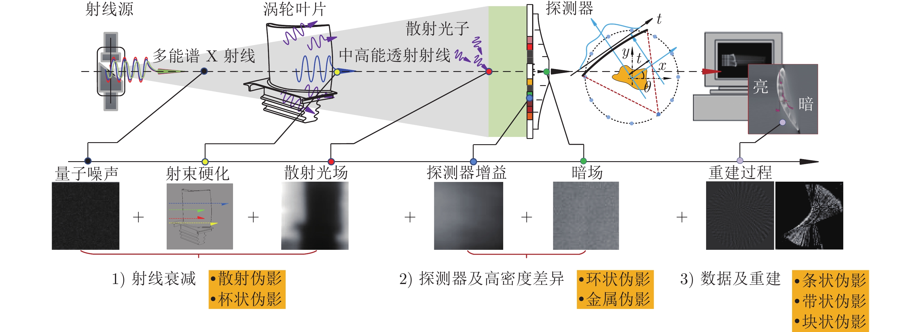
摘要: X射线工业计算机断层(Computerized tomography, CT)技术是一种先进的非接触式无损三维检测技术, 能在无损伤情况下以灰度图像的形式对物体内部结构进行全面、详细地分析, 在航空航天、工业生产、安检等领域发挥着重要的作用. 针对工业CT伪影严重降低图像质量问题, 对工业CT成像过程复杂伪影形成机理进行分析, 对不同类型伪影抑制方法进行归纳总结. 阐述了基于射线衰减、探测器及高密度差异、采样数据及重建等不同过程伪影成因及伪影消除相关算法的最新技术进展, 并对近年来人工智能深度学习背景下新兴的基于深度学习及神经网络的工业CT无损检测研究与发展方向进行了总结和展望.Abstract: Computerized tomography (CT) technology is an advanced non-destructive three-dimensional detection technology that can completely investigate the interior structure of an item in the form of grayscale images without contacting. It plays an important role in aerospace, industrial production, security inspection and other fields. Because industrial CT artifacts have had a significant impact on image resolution, this study examines the mechanism of coupling artifacts in the industrial CT imaging process, as well as the most recent technical advancements in artifact suppression methods. For the artifacts created during the ray attenuation process, the item containing scattering artifacts and cupping artifacts is described first. Second, the related technologies for artifact generated by the detector and the object suppression, such as ring artifacts and metal artifacts are discussed. And then, the latest developments in the suppression of streaking artifacts, banding artifacts and blocking artifacts are presented for the accessories generated by sampling data and reconstruction methods. By reviewing the research of industrial CT artifact suppression in recent years, the challenges and the development in the field of high-resolution imaging based on artificial intelligence in deep learning are summarized and prospected.
-
Key words:
- Industrial CT /
- artifact suppression /
- deep learning /
- intelligent detection
-
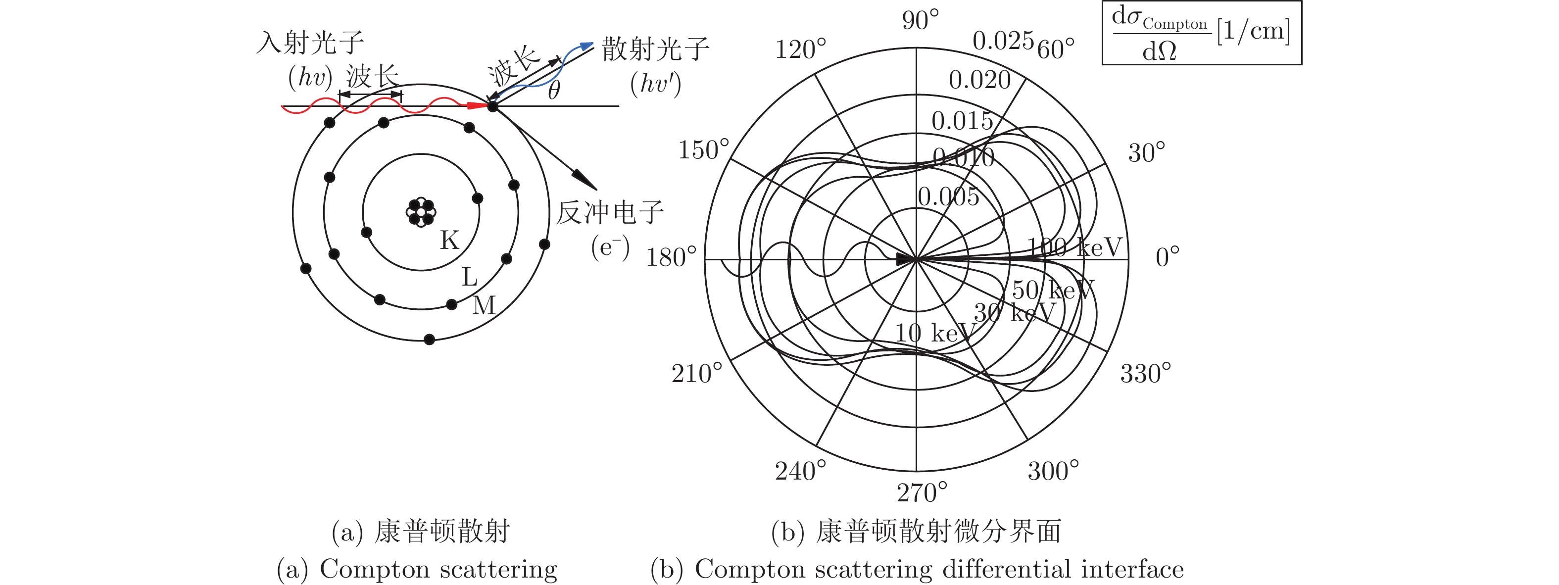
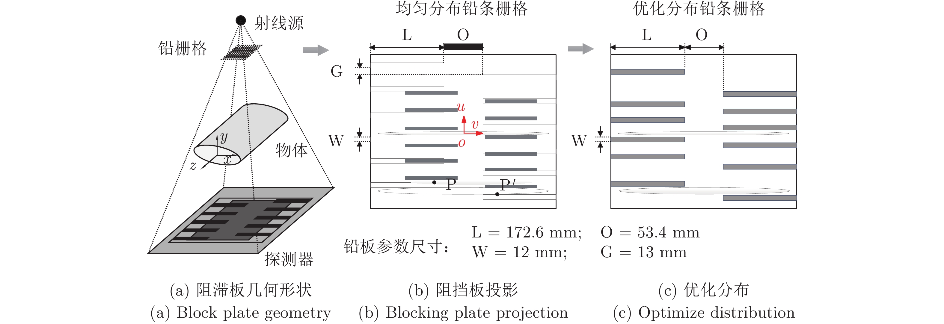
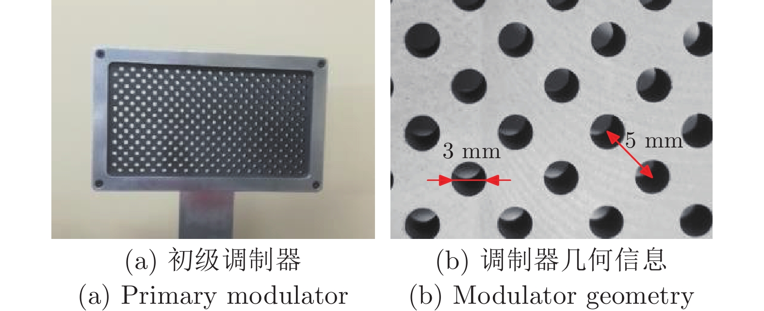
图 5 均匀分布和优化后光束阻滞示意图
Fig. 5 Schematic diagram of the beam block after uniform distribution and optimization
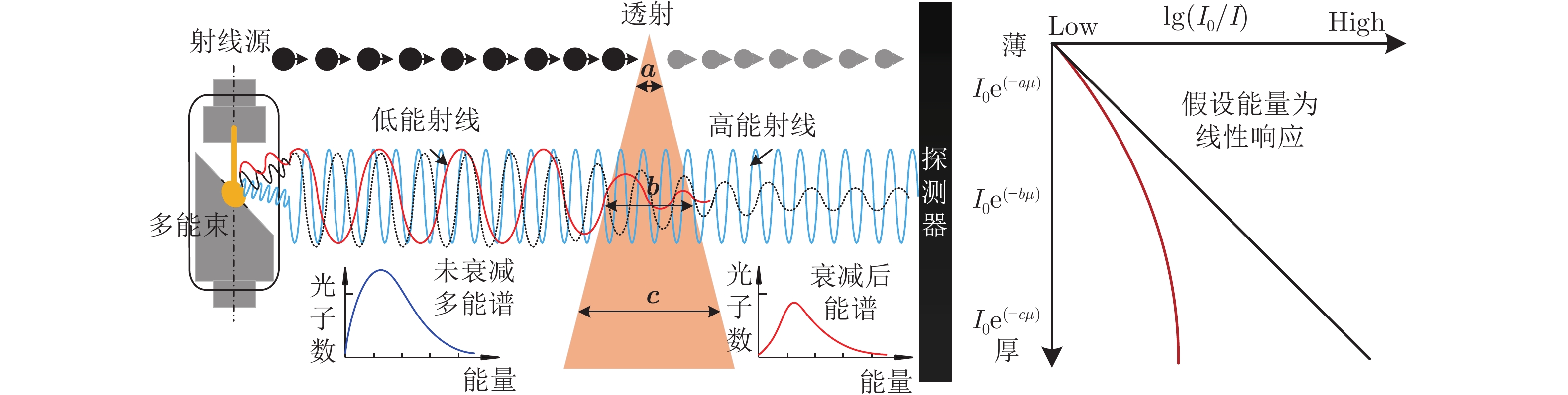
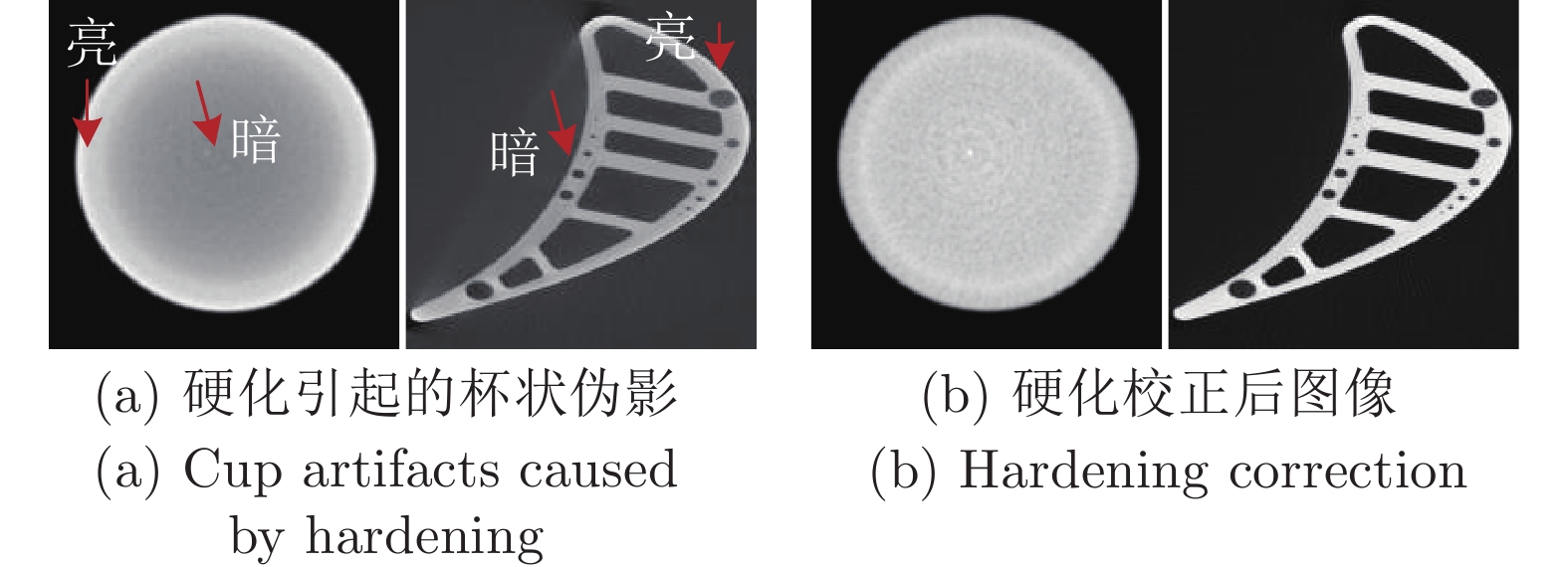
图 9 多能谱衰减过程产生硬化射束
Fig. 9 Hardened beam produced by multi-energy spectral attenuation process
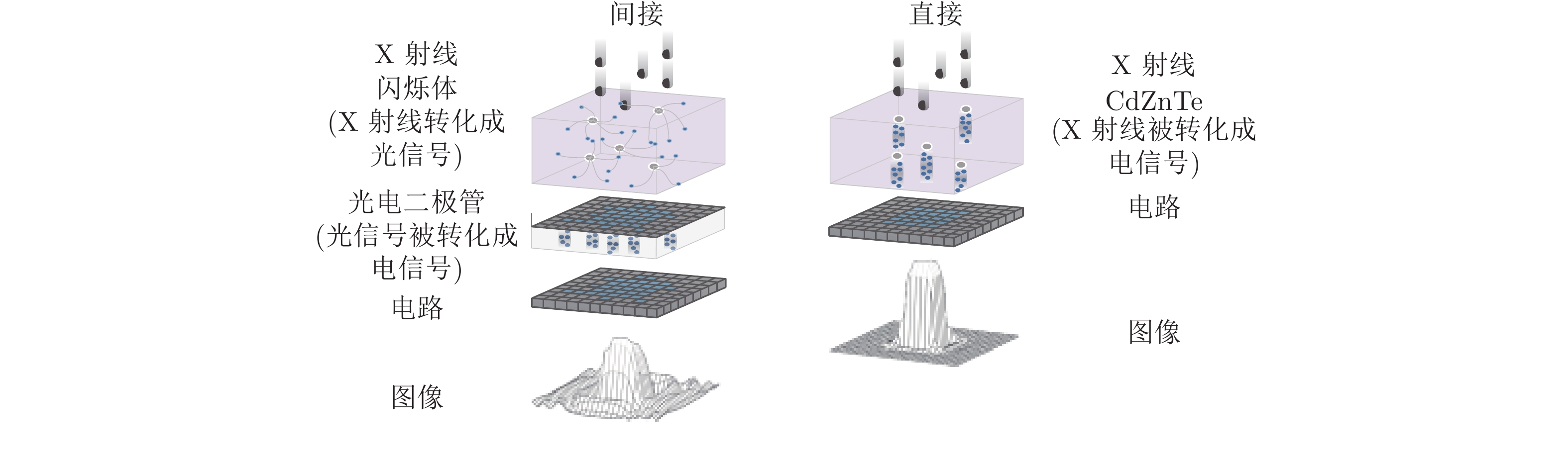
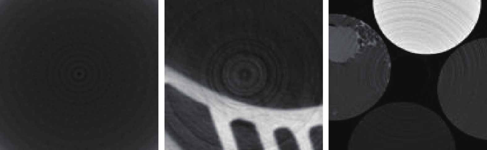
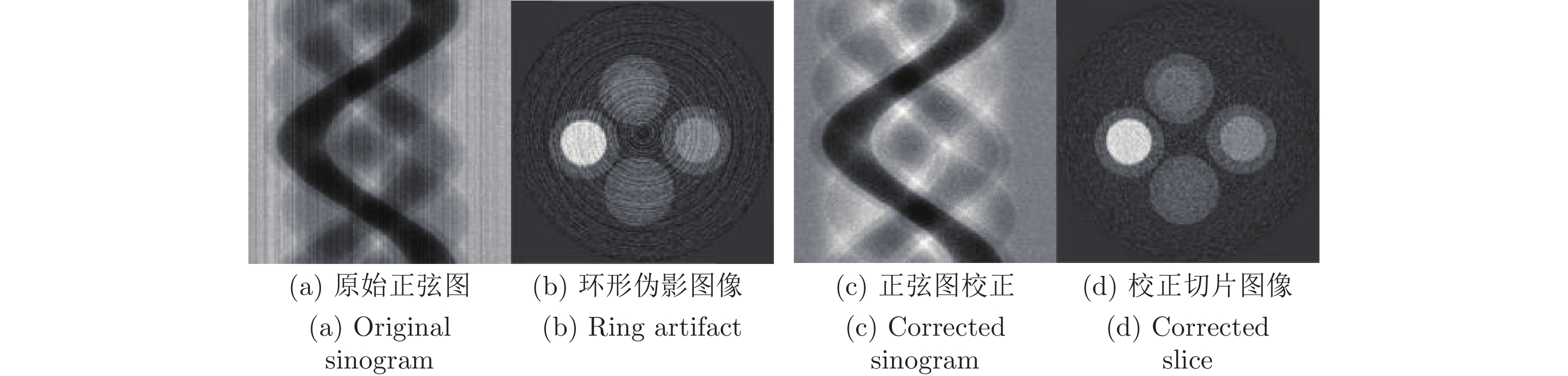
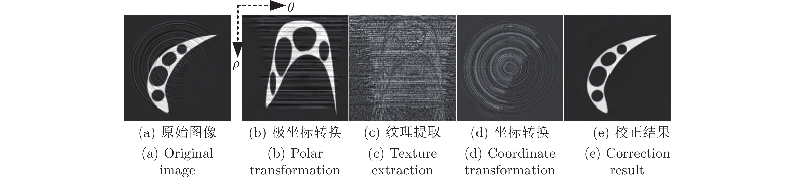
图 13 不同物体重建切片环形伪影示意图
Fig. 13 Schematic diagram of ring artifacts in reconstructed slices of different objects

图 17 条件生成对抗网络金属伪影校正流程
Fig. 17 Conditional generative adversarial network metal artifact correction process
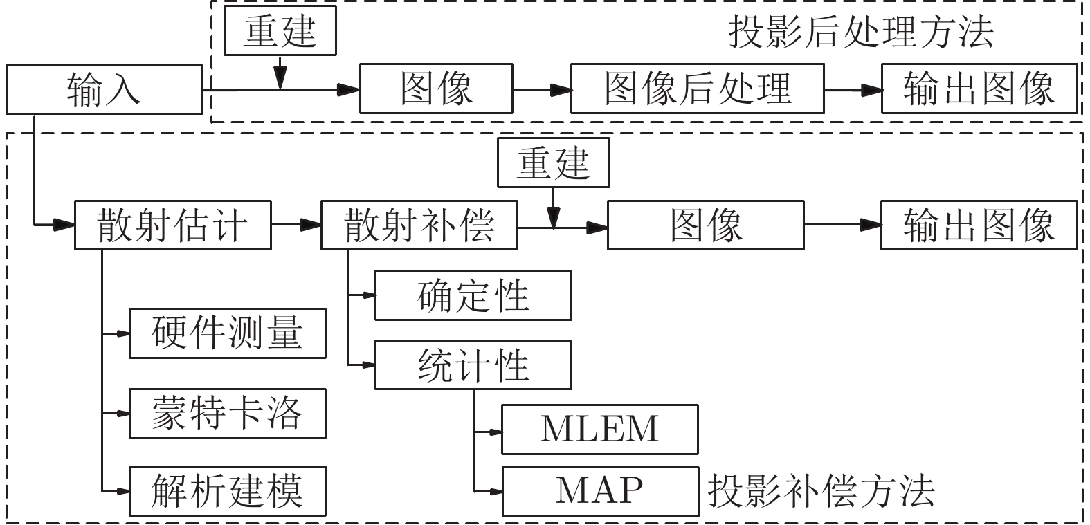
表 1 不同伪影的表现和产生原因及对应特征示意图
Table 1 Types of manifestations and causes corresponding to the characteristics of different artifacts
类型 成因 影响因素 特征 示例 散射伪影 射线强度空间频率较低,
散射光子干扰探测器接收到的光子并不
全是初始光子, 还包括散射
光子偏振干扰图像出现模糊, 边界
出现质量退化
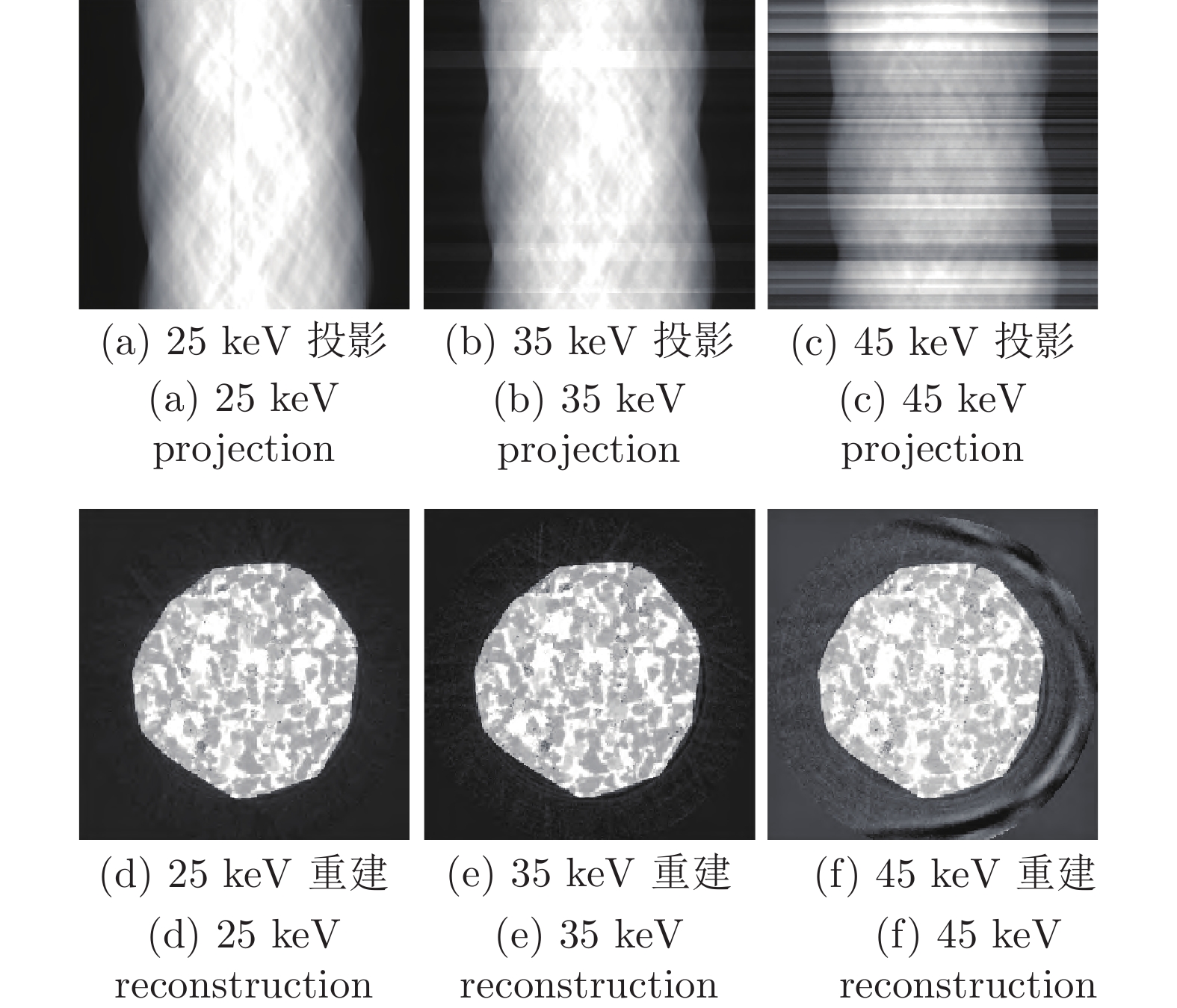
硬化伪影 (杯状伪影) 射线能谱发生变化,
射线光子吸收不均衡,
高能射线比重较大不同密度材料对射线能
量衰减程度不同图像出现外亮内暗的
灰度不均匀分布
杯状伪影 探测器受潮, 探测器不稳定 探测器的余晖效应、探测器
的响应不一致图像上出现圆圈状伪影环 
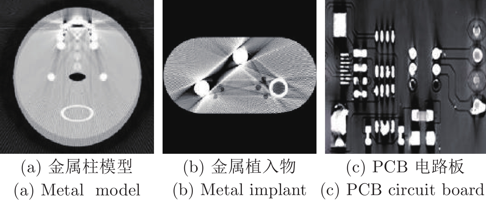
金属伪影 被检测物体中仅有单个金属 被扫描物中类似金属的
高衰减物质图像中呈现出明暗相间
的放射状伪影
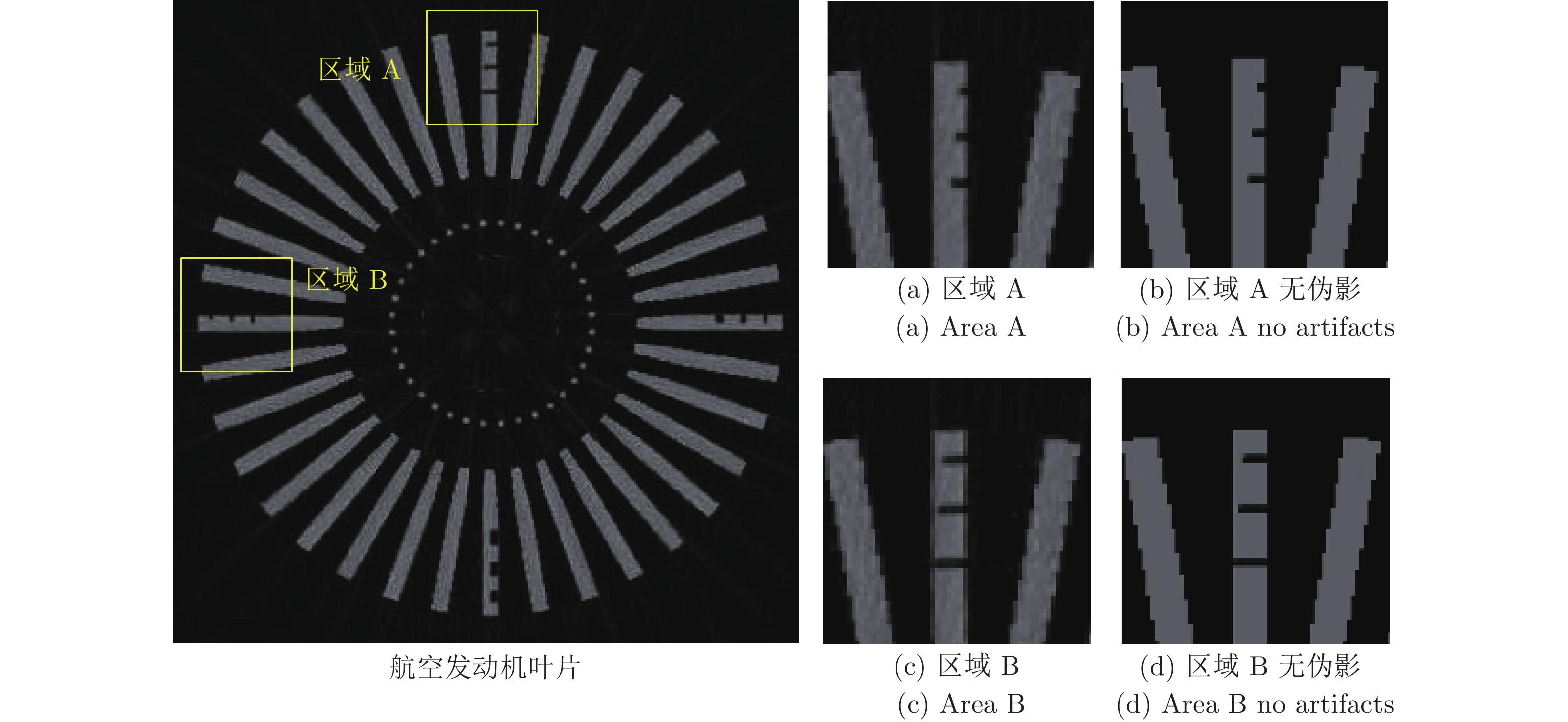
条状伪影 投影数据的不连续或中断 检测对象的移动和数据损失 重建图像存在线条状亮线区域 
带状伪影 光源的空间非均匀性 面源辐射波动性或光源不稳定性 图像局部偏亮或者偏暗 
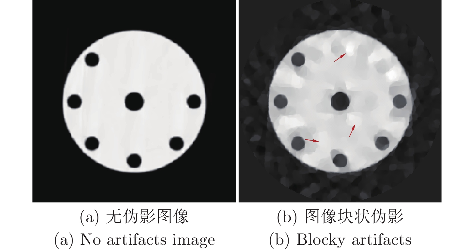
块状伪影 重建方法及数据结构 反映图像结构的字典训练不足 图像边缘细小结构扭曲 
 下载: 导出CSV
表 2 工业CT散射伪影抑制方法研究现状
Table 2 Research status of scattering artifact suppression methods for industrial CT
方法 主要贡献 实验结果 方法来源 主调制器掩模 补偿掩模影响的校正矩阵, 基于 B 样条曲线
的散射模型光谱 CT 对能谱先验信息依赖性比较强, 存在适应性问题 文献[38] 分段估计投影生成 提出一种最优的阻挡器分布, 以最小化缺失数据 将平均 CT 数误差从 86 个 Hounsfield 单位(HU) 减少到 9 HU, 并将图像对比度提高了 1.45 倍 文献[39] 增加距离减小散射 使用蒙特卡洛计算机模拟来计算散射投影比 (SPR) SPR 随着 X 射线能量的增加、材料密度的降低或 SID 的增加而降低 文献[40] 基于投影的等心和非等心成像法 构建了一个深度卷积自动编码器 (DCAE) 在非等中心患者 CT 采集中得到了成功运用 文献[48] 路径采样的散射估计 以规划 CT 图像的精确 CT 值作为先验信息, 自动控制每个粒子路径, 最终加速收敛 图像对比度提高, 散射伪影消除, 但是大量光子在传输过程中无法到达探测器, 使得估算不准确 文献[33] 卷积神经网络的散射校正 将深度卷积神经网络 (DCNN) 和残差学习框架 (RLF) 相结合 与没有 RLF 的网络相比, 所提出的方法具有更高的散射校正精度 文献[51]
下载: 导出CSV
表 3 工业CT硬化伪影/杯状伪影抑制方法研究现状
Table 3 Research status of cupping artifact suppression methods for industrial CT
方法 主要贡献 实验结果 方法来源 投影数据一致性条件约束 通过最小化一组投影对的不一致性, 迭代估计用于减少伪影的最佳多项式系数 减少了其他物理测量和几何误差对模型系数的干扰, 不需要校准也不需要先验信息 文献[61] 一种多项式射束硬化校正 利用三项式拟合构造一种多色投影模型, 并应用该模型来逼近实际投影数据 该模型能够有效地去除 X 射线硬化伪影, 但对于高密度物体往往效果有限, 且多项式系数获取过程复杂, 计算效率低 文献[63] 基于泰勒公式的曲线补偿 提出了一种获取光线穿过二值图像长度的方法, 构建了一种新的加权补偿校正模型 多色投影的伪影得到了有效的抑制, 该算法有望在工业无损检测中得到应用 文献[66] 基于光子计数探测器硬件 使用基于能量判别的 PCD 可以从本质上减少散射和射束硬化对图像质量的影响 与传统探测器相比, 能够在减少散射和波束硬化方面改善CT图像质量 文献[69]
下载: 导出CSV
表 4 工业CT金属伪影抑制方法研究现状
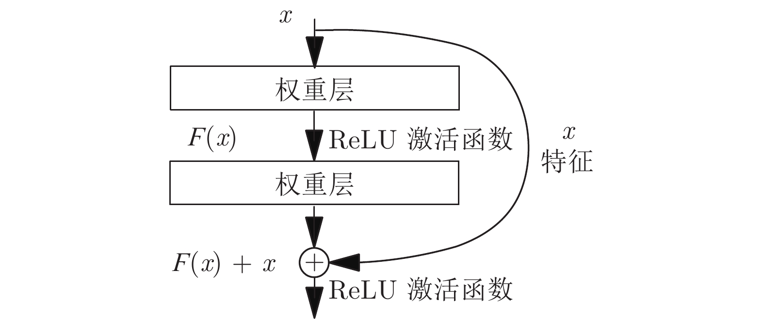
Table 4 Research status of metal artifact suppression methods for industrial CT
方法 主要贡献 实验结果 方法来源 基于投影校正 建立对金属区域投影值的校正模型, 采用单纯
形法迭代求解熵最小对多金属伪影的校正起到了良好的效果, 且校正后的图像质量优于插值校正法 文献[83] 基于先验图像校正 获得不含金属信息的先验图像, 后将先验数据与含金属投影进行插值 校正图像均方根误差最小、峰值信噪比最大, 保留图像边缘的同时, 可有效地抑制金属伪影 文献[85] 基于局部模型迭代校正 描述了一种将重建体自动划分为金属和非金属区域的方法 与常规重建相比, 该方案可使金属内部的硬化杯状伪影更少 文献[86] 基于残差编解码网络、混合GAN网络校正 利用投影数据开发了一种混合生成对抗网络
(GANs)的新组合掩模金字塔网络解决金属伪影校正研究中伪影消除不彻底、组织结构缺失等问题, 与传统重建算法相比, 结合迁移学习提高了学习网络的泛化性能 文献[88, 90]
下载: 导出CSV
-
[1] 戚俊成, 刘宾, 陈荣昌, 夏正德, 肖体乔. X射线光场成像技术研究. 物理学报, 2019, 68(2): Article No. 024202 doi: 10.7498/aps.68.20181555Qi Jun-Cheng, Liu Bin, Chen Rong-Chang, Xia Zheng-De, Xiao Ti-Qiao. X-ray three-dimensional imaging based on light field imaging technology. Acta Physica Sinica, 2019, 68(2): Article No. 024202 doi: 10.7498/aps.68.20181555 [2] 王林元, 刘宏奎, 李磊, 闫镔, 张瀚铭, 蔡爱龙, 等. 基于稀疏优化的计算机断层成像图像不完全角度重建综述. 物理学报, 2014, 63(20): Article No. 208702 doi: 10.7498/aps.63.208702Wang Lin-Yuan, Liu Hong-Kui, Li Lei, Yan Bin, Zhang Han-Ming, Cai Ai-Long, et al. Review of sparse optimization-based computed tomography image reconstruction from few-view projections. Acta Physica Sinica, 2014, 63(20): Article No. 208702 doi: 10.7498/aps.63.208702 [3] Jin S, Liu C H, Lai X M, Li F, He B. Bayesian network approach for ceramic shell deformation fault diagnosis in the investment casting process. The International Journal of Advanced Manufacturing Technology, 2017, 88(1-4): 663-674 doi: 10.1007/s00170-016-8795-x [4] du Plessis A, Rossouw P. X-ray computed tomography of a titanium aerospace investment casting. Case Studies in Nondestructive Testing and Evaluation, 2015, 3: 21-26 doi: 10.1016/j.csndt.2015.03.001 [5] Gameros A, De Chiffre L, Siller H R, Hiller J, Genta G. A reverse engineering methodology for nickel alloy turbine blades with internal features. CIRP Journal of Manufacturing Science and Technology, 2015, 9: 116-124 doi: 10.1016/j.cirpj.2014.12.001 [6] Li B, Chen L, Wang Y G, Gao M Q. 3D detection of internal defects for gas turbine blades. Insight-Non-Destructive Testing and Condition Monitoring, 2017, 59(7): 364-370 doi: 10.1784/insi.2017.59.7.364 [7] Przysowa R, Chalimoniuk M, Grzelka-Gajek D, Shakalo R, Karpenko A. CT inspection of cooled turbine blades. Journal of KONBiN, 2020, 50(3): 307-331 doi: 10.2478/jok-2020-0064 [8] 张辉, 张邹铨, 陈煜嵘, 吴天月, 钟杭, 王耀南. 工业铸件缺陷无损检测技术的应用进展与展望. 自动化学报, 2022, 48(4): 935-956Zhang Hui, Zhang Zou-Quan, Chen Yu-Rong, Wu Tian-Yue, Zhong Hang, Wang Yao-Nan. Application advance and prospect of nondestructive testing technology for industrial casting defects. Acta Automatica Sinica, 2022, 48(4): 935-956 [9] 朱雄泳, 吴炆芳, 陆许明, 谭洪舟, 邹兵兵. 基于一致性敏感哈希块匹配的HDR图像去伪影融合方法. 自动化学报, 2020, 46(7): 1496-1506Zhu Xiong-Yong, Wu Wen-Fang, Lu Xu-Ming, Tan Hong-Zhou, Zou Bing-Bing. High-dynamic-range image de-ghosting fusion method based on coherency sensitive hashing patch-match. Acta Automatica Sinica, 2020, 46(7): 1496-1506 [10] 李阳, 赵于前, 廖苗, 廖胜辉, 杨振. 基于水平集和形状描述符的腹部CT序列肝脏自动分割. 自动化学报, 2021, 47(2): 327-337Li Yang, Zhao Yu-Qian, Liao Miao, Liao Sheng-Hui, Yang Zhen. Automatic liver segmentation from CT volumes based on level set and shape descriptor. Acta Automatica Sinica, 2021, 47(2): 327-337 [11] 张朋, 张慧滔, 赵云松. X射线CT成像的数学模型及其有关问题. 数学建模及其应用, 2012, 1(1): 1-12 doi: 10.3969/j.issn.2095-3070.2012.01.001Zhang Peng, Zhang Hui-Tao, Zhao Yun-Song. The mathematical models on X-ray CT and their related issues. Mathematical Modeling and its Applications, 2012, 1(1): 1-12 doi: 10.3969/j.issn.2095-3070.2012.01.001 [12] Yang F Q, Zhang D H, Zhang H, Huang K D, Du Y, Teng M X. Streaking artifacts suppression for cone-beam computed tomography with the residual learning in neural network. Neurocomputing, 2020, 378: 65-78 doi: 10.1016/j.neucom.2019.09.087 [13] 袁翠云, 齐宏亮, 陈梓嘉, 吴书裕, 徐圆, 周凌宏. 基于投影域校正的CT图像环形伪影去除方法. 计算机工程与设计, 2017, 38(3): 735-738Yuan Cui-Yun, Qi Hong-Liang, Chen Zi-Jia, Wu Shu-Yu, Xu Yuan, Zhou Ling-Hong. CT image ring artifact reduction based on projection correction. Computer Engineering and Design, 2017, 38(3): 735-738 [14] Hu D L, Liu J, Lv T L, Zhao Q L, Zhang Y K, Quan G T, et al. Hybrid-domain neural network processing for sparse-view CT reconstruction. IEEE Transactions on Radiation and Plasma Medical Sciences, 2021, 5(1): 88-98 doi: 10.1109/TRPMS.2020.3011413 [15] Xu M R, Hu D L, Luo F L, Liu F L, Wang S Y, Wu W W. Limited-angle X-ray CT reconstruction using Image gradient ℓ_0-norm with dictionary learning. IEEE Transactions on Radiation and Plasma Medical Sciences, 2021, 5(1): 78-87 doi: 10.1109/TRPMS.2020.2991887 [16] 沈康, 刘松德, 施钧辉, 田超. 基于双域神经网络的稀疏视角光声图像重建. 中国激光, 2022, 49(5): Article No. 0507017Shen Kang, Liu Song-De, Shi Jun-Hui, Tian Chao. Dual-domain neural network for sparse-view photoacoustic image reconstruction. Chinese Journal of Lasers, 2022, 49(5): Article No. 0507017 [17] Greffier J, Frandon J, Larbi A, Beregi J P, Pereira J. CT iterative reconstruction algorithms: A task-based image quality assessment. European Radiology, 2020, 30(1): 487-500 doi: 10.1007/s00330-019-06359-6 [18] 胡伟达, 李庆, 陈效双, 陆卫. 具有变革性特征的红外光电探测器. 物理学报, 2019, 68(12): Article No. 120701 doi: 10.7498/aps.68.20190281Hu Wei-Da, Li Qing, Chen Xiao-Shuang, Lu Wei. Recent progress on advanced infrared photodetectors. Acta Physica Sinica, 2019, 68(12): Article No. 120701 doi: 10.7498/aps.68.20190281 [19] 沈百飞, 吉亮亮, 张晓梅, 步志刚, 徐建彩. 强场X射线激光物理. 物理学报, 2021, 70(8): Article No. 084101Shen Bai-Fei, Ji Liang-Liang, Zhang Xiao-Mei, Bu Zhi-Gang, Xu Jian-Cai. High field X-ray laser physics. Acta Physica Sinica, 2021, 70(8): Article No. 084101 [20] Kak A C, Slaney M, Wang G. Principles of computerized tomographic imaging. Medical Physics, 2002, 29(1): Article No. 107 [21] Ito S, Toda N. Improvement of CT reconstruction using scattered X-rays. IEICE Transactions on Information and Systems, 2021, E104.D(8): 1378-1385 doi: 10.1587/transinf.2020EDP7241 [22] Wiegert D I J, Ohm U D I J R. Scattered Radiation in Cone-beam Computed Tomography: Analysis, Quantification and Compensation. Germany: Publication Server of RWTH Aachen University, 2007. 12−19 [23] 刘建邦, 席晓琦, 韩玉, 李磊, 卜海兵, 闫镔. 基于K-N模型的锥束CT散射伪影校正方法. 光学学报, 2018, 38(11): Article No. 1134001 doi: 10.3788/AOS201838.1134001Liu Jian-Bang, Xi Xiao-Qi, Han Yu, Li Lei, Bu Hai-Bing, Yan Bin. A new scattering artifact correction method based on K-N formula for cone-beam computed tomography. Acta Optica Sinica, 2018, 38(11): Article No. 1134001 doi: 10.3788/AOS201838.1134001 [24] 张定华, 胡栋材, 黄魁东, 孔永茂. 基于射束衰减网格的锥束CT散射校正方法. 中国机械工程, 2009, 20(6): 639-643 doi: 10.3321/j.issn:1004-132X.2009.06.003Zhang Ding-Hua, Hu Dong-Cai, Huang Kui-Dong, Kong Yong-Mao. Beam attenuation grid-based scatter correction method for cone beam CT. China Mechanical Engineering, 2009, 20(6): 639-643 doi: 10.3321/j.issn:1004-132X.2009.06.003 [25] 胡栋材, 陈浩, 张定华. 基于平板探测器的锥束CT散射校正方法. CT理论与应用研究, 2009, 18(1): 16-22Hu Dong-Cai, Chen Hao, Zhang Ding-Hua. Scatter correction method for flat-panel detector-based cone beam CT. CT Theory and Applications, 2009, 18(1): 16-22 [26] Bootsma G J, Verhaegen F, Jaffray D A. The effects of compensator and imaging geometry on the distribution of X-ray scatter in CBCT. Medical Physics, 2011, 38(2): 897-914 doi: 10.1118/1.3539575 [27] 谢世朋, 罗立民. 基于衰减板的锥束CT散射校正. 电子学报, 2011, 39(7): 1708-1711Xie Shi-Peng, Luo Li-Min. Scatter correction for cone beam CT using attenuation baffle. Acta Electronica Sinica, 2011, 39(7): 1708-1711 [28] Maier J, Sawall S, Knaup M, Kachelrieß M. Deep scatter estimation (DSE): Accurate real-time scatter estimation for X-ray CT using a deep convolutional neural network. Journal of Nondestructive Evaluation, 2018, 37(3): Article No. 57 doi: 10.1007/s10921-018-0507-z [29] Zhou X, Sun J F, Jiang P, Liu D, Wang Q. Effect of optical-scattering characteristics with modified kernel-based model on the triggering probability of Gm-APD. Journal of Quantitative Spectroscopy and Radiative Transfer, 2020, 243: Article No. 106814 doi: 10.1016/j.jqsrt.2019.106814 [30] Ouyang L, Song K, Wang J. A moving blocker system for cone-beam computed tomography scatter correction. Medical Physics, 2013, 40(7): Article No. 071903 [31] Ritschl L, Fahrig R, Knaup M, Maier J, Kachelrieß M. Robust primary modulation-based scatter estimation for cone-beam CT. Medical Physics, 2015, 42(1): 469-478 doi: 10.1118/1.4903261 [32] Bier B, Berger M, Maier A, Kachelrieß M, Ritschl L, Müller K, et al. Scatter correction using a primary modulator on a clinical angiography C-arm CT system. Medical Physics, 2017, 44(9): e125-e137 doi: 10.1002/mp.12094 [33] Zhang Y M, Chen Y S, Zhong A N. Jia X, Wu S Y, Qi H L, et al. Scatter correction based on adaptive photon path-based Monte Carlo simulation method in multi-GPU platform. Computer Methods and Programs in Biomedicine, 2020, 194: Article No. 105487 doi: 10.1016/j.cmpb.2020.105487 [34] Xiang H W, Lim H, Fessler J A, Dewaraja Y K. A deep neural network for fast and accurate scatter estimation in quantitative SPECT/CT under challenging scatter conditions. European Journal of Nuclear Medicine and Molecular Imaging, 2020, 47(13): 2956-2967 doi: 10.1007/s00259-020-04840-9 [35] Iskender B, Bresler Y. A physics-motivated DNN for X-ray CT scatter correction. In: Proceedings of the 17th International Symposium on Biomedical Imaging. Iowa, USA: IEEE, 2019. 609−613 [36] Tien H J, Yang H C, Shueng P W, Chen J C. Cone-beam CT image quality improvement using Cycle-Deblur consistent adversarial networks (Cycle-Deblur GAN) for chest CT imaging in breast cancer patients. Scientific Reports, 2021, 11(1): Article No. 1133 doi: 10.1038/s41598-020-80803-2 [37] Liang X K, Li N, Zhang Z C, Yu S D, Qin W J, Li Y F, et al. Shading correction for volumetric CT using deep convolutional neural network and adaptive filter. Quantitative Imaging in Medicine and Surgery, 2019, 9(7): 1242-1254 doi: 10.21037/qims.2019.05.19 [38] Pivot O, Fournier C, Tabary J, Letang J M, Rit S. Scatter correction for spectral CT using a primary modulator mask. IEEE Transactions on Medical Imaging, 2020, 39(6): 2267-2276 doi: 10.1109/TMI.2020.2970296 [39] Liang X K, Jiang Y K, Zhao W, Zhang Z C, Luo C, Xiong J, et al. Scatter correction for a clinical cone-beam CT system using an optimized stationary beam blocker in a single scan. Medical physics, 2019, 46(7): 3165-3179 doi: 10.1002/mp.13568 [40] Tu S J. Scatter-to-primary ratio in cone beam computed tomography with extended source to image-receptor distance for image-guided proton beam therapy system. Radiation Physics and Chemistry, 2020, 170: Article No. 108667 doi: 10.1016/j.radphyschem.2019.108667 [41] Siewerdsen J H, Moseley D J, Bakhtiar B, Richard S, Jaffray D A. The influence of antiscatter grids on soft-tissue detectability in cone-beam computed tomography with flat-panel detectors. Medical Physics, 2004, 31(12): 3506-3520 doi: 10.1118/1.1819789 [42] Yang F Q, Zhang D H, Zhang H, Huang K D. Scattering measurement and estimation in angular sequence for cone-beam CT based on projection structural tensor and modeling. Journal of X-Ray Science and Technology, 2019, 27(5): 965-979 doi: 10.3233/XST-190528 [43] Yang F Q, Zhang D H, Huang K D, Shi W L, Wang X Y. Scattering estimation for cone-beam CT using local measurement based on compressed sensing. IEEE Transactions on Nuclear Science, 2018, 65(3): 941-949 doi: 10.1109/TNS.2018.2803739 [44] Sun M, Star-Lack J M. Improved scatter correction using adaptive scatter kernel superposition. Physics in Medicine & Biology, 2010, 55(22): 6695-6720 [45] Bootsma G J, Verhaegen F, Jaffray D A. Efficient scatter distribution estimation and correction in CBCT using concurrent Monte Carlo fitting. Medical Physics, 2015, 42(1): 54-68 [46] Yan H, Mou X Q, Tang S J, Xu Q, Zankl M. Projection correlation based view interpolation for cone beam CT: Primary fluence restoration in scatter measurement with a moving beam stop array. Physics in Medicine & Biology, 2010, 55(21): 6353-6375 [47] 戎军艳, 刘文磊, 高鹏, 廖琪梅, 卢虹冰. 锥束CT散射抑制方法综述. CT理论与应用研究, 2016, 25(2): 235-250 doi: 10.15953/j.1004-4140.2016.25.02.15Rong Jun-Yan, Liu Wen-Lei, Gao Peng, Liao Qi-Mei, Lu Hong-Bing. The review of scatter suppression methods in cone beam CT. CT Theory and Applications, 2016, 25(2): 235-250 doi: 10.15953/j.1004-4140.2016.25.02.15 [48] van der Heyden B, Uray M, Fonseca G P, Huber P, Us D, Messner I, et al. A Monte Carlo based scatter removal method for non-isocentric cone-beam CT acquisitions using a deep convolutional autoencoder. Physics in Medicine & Biology, 2020, 65(14): Article No. 145002 [49] 施俊, 汪琳琳, 王珊珊, 陈艳霞, 王乾, 魏冬铭, 等. 深度学习在医学影像中的应用综述. 中国图象图形学报, 2020, 25(10): 1953-1981 doi: 10.11834/jig.200255Shi Jun, Wang Lin-Lin, Wang Shan-Shan, Chen Yan-Xia, Wang Qian, Wei Dong-Ming, et al. Applications of deep learning in medical imaging: A survey. Journal of Image and Graphics, 2020, 25(10): 1953-1981 doi: 10.11834/jig.200255 [50] He K M, Zhang X Y, Ren S Q, Sun J. Deep residual learning for image recognition. In: Proceedings of the IEEE Conference on Computer Vision and Pattern Recognition (CVPR). Las Vegas, NV, USA: IEEE, 2016. 770−778 [51] Jiang Y K, Yang C L, Yang P F, Hu X, Luo C, Xue Y, et al. Scatter correction of cone-beam CT using a deep residual convolution neural network (DRCNN). Physics in Medicine & Biology, 2019, 64(14): Article No. 145003 [52] Hansen D C, Landry G, Kamp F, Li M L, Belka C, Parodi K, et al. ScatterNet: A convolutional neural network for cone-beam CT intensity correction. Medical Physics, 2018, 45(11): 4916-4926 doi: 10.1002/mp.13175 [53] Nomura Y, Xu Q, Shirato H, Shimizu S, Xing L. Projection-domain scatter correction for cone beam computed tomography using a residual convolutional neural network. Medical Physics, 2019, 46(7): 3142-3155 doi: 10.1002/mp.13583 [54] Cao W C, Hawker S, Fardell G, Price B, Dewulf W. An improved segmentation method for multi-material beam hardening correction in industrial X-ray computed tomography. Measurement Science and Technology, 2019, 30(12): Article No. 125403 doi: 10.1088/1361-6501/ab30bb [55] Tang S J, Huang K D, Cheng Y Y, Mou X Q, Tang X Y. Optimization based beam-hardening correction in CT under data integral invariant constraint. Physics in Medicine & Biology, 2018, 63(13): Article No. 135015 [56] Sarkar S, Wahi P, Munshi P. An empirical correction method for beam-hardening artifact in computerized tomography (CT) images. NDT & E International, 2019, 102: 104-113 [57] Ahmed O M H, Song Y S. A review of common beam hardening correction methods for industrial X-ray computed tomography. Sains Malaysiana, 2018, 47(8): 1883-1890 doi: 10.17576/jsm-2018-4708-29 [58] Yang Q, Fullagar W K, Myers G R, Latham S J, Varslot T, Sheppard A P, et al. X-ray attenuation models to account for beam hardening in computed tomography. Applied Optics, 2020, 59(29): 9126-9136 doi: 10.1364/AO.402304 [59] Shi H L, Yang Z, Luo S Q. Reduce beam hardening artifacts of polychromatic X-ray computed tomography by an iterative approximation approach. Journal of X-Ray Science and Technology, 2017, 25(3): 417-428 doi: 10.3233/XST-16187 [60] Luo S H, Wu H Z, Sun Y, Li J, Li G, Gu N. A fast beam hardening correction method incorporated in a filtered back-projection based MAP algorithm. Physics in Medicine & Biology, 2017, 62(5): 1810-1830 [61] Abdurahman S, Frysch R, Bismark R, Melnik S, Beuing O, Rose G. Beam hardening correction using cone beam consistency conditions. IEEE Transactions on Medical Imaging, 2018, 37(10): 2266-2277 doi: 10.1109/TMI.2018.2840343 [62] Levi J, Wu H, Eck B L, Fahmi R, Vembar M, Dhanantwar A, et al. Comparison of automated beam hardening correction (ABHC) algorithms for myocardial perfusion imaging using computed tomography. Medical Physics, 2021, 48(1): 287-299 doi: 10.1002/mp.14599 [63] Xiu G Y, Yuan C Y, Chen X H, Li X S. An innovative beam hardening correction method for computed tomography systems. Traitement du Signal, 2019, 36(6): 515-520 doi: 10.18280/ts.360606 [64] Ahmed O M H, Song Y S, Xie Z Y, Ayoub A A T. Calculation of beam hardening in industrial X-ray computed tomography and its correction using filtration and linearization methods. IOP Conference Series: Earth and Environmental Science, 2020, 461: Article No. 012082 [65] Lifton J J. Multi-material linearization beam hardening correction for computed tomography. Journal of X-Ray Science and Technology, 2017, 25(4): 629−640 [66] Yang F Q, Zhang D H, Zhang H, Huang K D. Cupping artifacts correction for polychromatic X-ray cone-beam computed tomography based on projection compensation and hardening behavior. Biomedical Signal Processing and Control, 2020, 57: Article No. 101823 doi: 10.1016/j.bspc.2019.101823 [67] Bismark R N K, Frysch R, Abdurahman S, Beuing O, Blessing M, Rose G. Reduction of beam hardening artifacts on real C-arm CT data using polychromatic statistical image reconstruction. Zeitschrift Für Medizinische Physik, 2020, 30(1): 40-50 [68] Abella M, Martínez C, Desco M, Vaquero J J, Fessler J A. Simplified statistical image reconstruction for X-ray CT with beam-hardening artifact compensation. IEEE Transactions on Medical Imaging, 2020, 39(1): 111-118 doi: 10.1109/TMI.2019.2921929 [69] Schumacher D, Sharma R, Grager J C, Schrapp M. Scatter and beam hardening reduction in industrial computed tomography using photon counting detectors. Measurement Science and Technology, 2018, 29(7): Article No. 075101 [70] Kimoto N, Hayashi H, Asakawa T, Lee C, Asahara T, Maeda T, et al. Effective atomic number image determination with an energy-resolving photon-counting detector using polychromatic X-ray attenuation by correcting for the beam hardening effect and detector response. Applied Radiation and Isotopes, 2021, 170: Article No. 109617 doi: 10.1016/j.apradiso.2021.109617 [71] Watanabe M, Sato E, Yoshida S, Yoshioka K, Oda Y, Moriyama H, et al. Photon-counting X-ray computed tomography using a YAP(Ce)-PMT detector and beam hardening. In: Proceedings of SPIE 11114, Hard X-Ray, Gamma-Ray, and Neutron Detector Physics XXI. San Diego, USA: SPIE, 2019. Article No. 111141D [72] Šalplachta J, Zikmund T, Zemek M, Břínek A, Takeda Y, Omote K, et al. Complete ring artifacts reduction procedure for lab-based X-ray nano CT systems. Sensors, 2021, 21(1): Article No. 238 [73] Sadi F, Lee S Y, Hasan K. Removal of ring artifacts in computed tomographic imaging using iterative center weighted median filter. Computers in Biology and Medicine, 2010, 40(1): 109-118 doi: 10.1016/j.compbiomed.2009.11.007 [74] Hasan K, Sadi F, Lee S Y. Removal of ring artifacts in micro-CT imaging using iterative morphological filters. Signal, Image and Video Processing, 2012, 6(1): 41-53 doi: 10.1007/s11760-010-0170-z [75] Prell D, Kyriakou Y, Kalender W A. Comparison of ring artifact correction methods for flat-detector CT. Physics in Medicine & Biology, 2009, 54(12): 3881-3895 [76] Wei Z P, Wiebe S, Chapman D. Ring artifacts removal from synchrotron CT image slices. Journal of Instrumentation, 2013, 8: Article No. C06006 [77] Anas E M A, Kim J G, Lee S Y, Hasan K. High-quality 3D correction of ring and radiant artifacts in flat panel detector-based cone beam volume CT imaging. Physics in Medicine & Biology, 2011, 56(19): 6495-6519 [78] Eldib M E, Hegazy M, Mun Y J, Cho M H, Cho M H, Lee S Y. A ring artifact correction method: Validation by micro-CT imaging with flat-panel detectors and a 2D photon-counting detector. Sensors, 2017, 17(2): Article No. 269 doi: 10.3390/s17020269 [79] 王珏, 黄苏红, 蔡玉芳. 工业CT图像环形伪影校正. 光学 精密工程, 2010, 18(5): 1226-1233Wang Jue, Huang Su-Hong, Cai Yu-Fang. Ring artifact correction for ICT images. Optics and Precision Engineering, 2010, 18(5): 1226-1233 [80] 霍其润, 李建武, 陆耀, 秦明. 基于变分的CT图像环形伪影校正. 自动化学报, 2019, 45(9): 1713-1726 doi: 10.16383/j.aas.c180258Huo Qi-Run, Li Jian-Wu, Lu Yao, Qin Ming. Variation-based ring artifact correction in CT images. Acta Automatica Sinica, 2019, 45(9): 1713-1726 doi: 10.16383/j.aas.c180258 [81] Yang Y F, Zhang D H, Yang F Q, Teng M X, Du Y, Huang K D. Post-processing method for the removal of mixed ring artifacts in CT images. Optics Express, 2020, 28(21): 30362-30378 doi: 10.1364/OE.401088 [82] 汪涛, 夏文军, 赵云松, 张意. CT金属伪影去除研究进展. 中国体视学与图像分析, 2020, 25(3): 207-223 doi: 10.13505/j.1007-1482.2020.25.03.001Wang Tao, Xia Wen-Jun, Zhao Yun-Song, Zhang Yi. Review of metal artifact reduction in computed tomography. Chinese Journal of Stereology and Image Analysis, 2020, 25(3): 207-223 doi: 10.13505/j.1007-1482.2020.25.03.001 [83] 魏星, 闫镔, 张峰, 李永丽, 席晓琦, 李磊. 多金属物体CT图像的金属伪影校正. 物理学报, 2014, 63(5): Article No. 058702Wei Xing, Yan Bin, Zhang Feng, Li Yong-Li, Xi Xiao-Qi, Li Lei. Reduction of metal artifacts caused by multiple metallic objects in computed tomography. Acta Physica Sinica, 2014, 63(5): Article No. 058702 [84] 李磊, 韩玉, 席晓琦, 王敬雨, 闫镔. 基于能谱滤波和图像残差重投影的CT图像金属伪影校正方法. 信息工程大学学报, 2017, 18(3): 288-293 doi: 10.3969/j.issn.1671-0673.2017.03.007Li Lei, Han Yu, Xi Xiao-Qi, Wang Jing-Yu, Yan Bin. Metal artifact correction method in computed tomography based on spectra filtering and image residual reprojection. Journal of Information Engineering University, 2017, 18(3): 288-293 doi: 10.3969/j.issn.1671-0673.2017.03.007 [85] 刘仰川, 高鹏, 朱叶晨, 高欣. 一种基于先验图像的锥束CT金属伪影校正算法. 图学学报, 2020, 41(4): 529-538Liu Yang-Chuan, Gao Peng, Zhu Ye-Chen, Gao Xin. A prior-image-based metal artifact reduction method for cone beam CT. Journal of Graphics, 2020, 41(4): 529-538 [86] Van Slambrouck K, Nuyts J. Metal artifact reduction in computed tomography using local models in an image block-iterative scheme. Medical Physics, 2012, 39(11): 7080-7093 doi: 10.1118/1.4762567 [87] Hegazy M A A, Cho M H, Cho M H, Lee S Y. U-net based metal segmentation on projection domain for metal artifact reduction in dental CT. Biomedical Engineering Letters, 2019, 9(3): 375-385 doi: 10.1007/s13534-019-00110-2 [88] 马燕, 余海军, 钟发生, 刘丰林. 基于残差编解码网络的CT图像金属伪影校正. 仪器仪表学报, 2020, 41(8): 160-169 doi: 10.19650/j.cnki.cjsi.J2006503Ma Yan, Yu Hai-Jun, Zhong Fa-Sheng, Liu Feng-Lin. CT metal artifact reduction based on the residual encoder-decoder network. Chinese Journal of Scientific Instrument, 2020, 41(8): 160-169 doi: 10.19650/j.cnki.cjsi.J2006503 [89] Zhang Y B, Yu H Y. Convolutional neural network based metal artifact reduction in X-ray computed tomography. IEEE Transactions on Medical Imaging, 2018, 37(6): 1370-1381 doi: 10.1109/TMI.2018.2823083 [90] Gomi T, Sakai R, Hara H, Watanabe Y, Mizukami S. Usefulness of a metal artifact reduction algorithm in digital tomosynthesis using a combination of hybrid generative adversarial networks. Diagnostics, 2021, 11(9): Article No. 1629 doi: 10.3390/diagnostics11091629 [91] Hao S L, Liu J, Chen Y, Liu B D, Wei C F, Zhu J, et al. A wavelet transform-based photon starvation artifacts suppression algorithm in CT imaging. Physics in Medicine & Biology, 2020, 65(23): Article No. 235039 [92] Yazdi M, Beaulieu L. Artifacts in spiral X-ray CT scanners: Problems and solutions. International Journal of Electrical, Computer, Energetic, Electronic and Communication Engineering, 2007, 1(11): 1599−1603 [93] 李保磊, 莫阳, 张萍宇, 李斌, 张耀军. X射线CT成像条状伪影校正. 原子能科学技术, 2017, 51(11): 2075-2078 doi: 10.7538/yzk.2017.youxian.0225Li Bao-Lei, Mo Yang, Zhang Ping-Yu, Li Bin, Zhang Yao-Jun. Streak artifact correction in X-ray CT imaging. Atomic Energy Science and Technology, 2017, 51(11): 2075-2078 doi: 10.7538/yzk.2017.youxian.0225 [94] Yang F Q, Zhang D H, Zhang H, Huang K D, Du Y. Fusion reconstruction algorithm to ill-posed projection (FRAiPP) for artifacts suppression on X-ray computed tomography. Nuclear Instruments and Methods in Physics Research Section A: Accelerators, Spectrometers, Detectors and Associated Equipment, 2020, 976: Article No. 164263 doi: 10.1016/j.nima.2020.164263 [95] 康慧, 高红霞, 胡跃明, 郭琪伟. 基于Bregman迭代的CT图像重建算法. 自动化学报, 2013, 39(9): 1570-1575Kang Hui, Gao Hong-Xia, Hu Yue-Ming, Guo Qi-Wei. Reconstruction algorithm based on Bregman iteration. Acta Automatica Sinica, 2013, 39(9): 1570-1575 [96] 王娜, 张权, 刘祎, 贾丽娜, 桂志国. 基于可变阶变分模型的医用低剂量CT图像去噪. 北京航空航天大学学报, 2019, 45(9): 1757-1764 doi: 10.13700/j.bh.1001-5965.2018.0775Wang Na, Zhang Quan, Liu Yi, Jia Li-Na, Gui Zhi-Guo. Medical low-dose CT image denoising based on variable order variational model. Journal of Beijing University of Aeronautics and Astronautics, 2019, 45(9): 1757-1764 doi: 10.13700/j.bh.1001-5965.2018.0775 [97] 卢洪义, 陈庆贵, 周红梅, 赵汝岩, 张宗伟, 喻舟. 固体火箭发动机CT图像条状伪影校正. 航空动力学报, 2016, 31(10): 2515-2521 doi: 10.13224/j.cnki.jasp.2016.10.027Lu Hong-Yi, Chen Qing-Gui, Zhou Hong-Mei, Zhao Ru-Yan, Zhang Zong-Wei, Yu Zhou. Streak artifacts correction of solid rocket motor's CT image. Journal of Aerospace Power, 2016, 31(10): 2515-2521 doi: 10.13224/j.cnki.jasp.2016.10.027 [98] Xu Q, Yu H Y, Mou X Q, Zhang L, Hsieh J, Wang G. Low-dose X-ray CT reconstruction via dictionary learning. IEEE Transactions on Medical Imaging, 2012, 31(9): 1682-1697 doi: 10.1109/TMI.2012.2195669 [99] Karimi D, Ward R. Reducing streak artifacts in computed tomography via sparse representation in coupled dictionaries. Medical Physics, 2016, 43(3): 1473-1486 doi: 10.1118/1.4942376 [100] Yang F Q, Zhang D H, Huang K D, Gao Z Z, Yang Y F. Incomplete projection reconstruction of computed tomography based on the modified discrete algebraic reconstruction technique. Measurement Science and Technology, 2018, 29(2): Article No. 025405 [101] Mori I, Machida Y, Osanai M, Iinuma K. Photon starvation artifacts of X-ray CT: Their true cause and a solution. Radiological Physics and Technology, 2013, 6(1): 130-141 doi: 10.1007/s12194-012-0179-9 [102] 孔慧华, 杨玉双. 同步辐射CT投影数据中的带状伪影及重建误差分析. 中北大学学报(自然科学版), 2016, 37(1): 61-66, 75Kong Hui-Hua, Yang Yu-Shuang. Analysis on reconstructed errors and band-artifacts in projections for synchrotron radiation computed tomography. Journal of North University of China (Natural Science Edition), 2016, 37(1): 61-66, 75 [103] Sun B Y, Hayakawa Y. Impact of statistical reconstruction and compressed sensing algorithms on projection data elimination during X-ray CT image reconstruction. Oral Radiology, 2018, 34(3): 237-244 doi: 10.1007/s11282-017-0308-6 [104] Sun L Y, Fan Z W, Fu X Y, Huang Y, Ding X H, Paisley J. A deep information sharing network for multi-contrast compressed sensing MRI reconstruction. IEEE Transactions on Image Processing, 2019, 28(12): 6141-6153 doi: 10.1109/TIP.2019.2925288 [105] Dinh K Q, Shim H J, Jeon B. Weighted overlapped recovery for blocking artefacts reduction in block-based compressive sensing of images. Electronics Letters, 2015, 51(1): 48-50 doi: 10.1049/el.2014.3200 [106] He G, Huang N J. A new particle swarm optimization algorithm with an application. Applied Mathematics and Computation, 2014, 232: 521-528 doi: 10.1016/j.amc.2014.01.028 [107] Singh A, Singh J. A content adaptive method of de-blocking and super-resolution of compressed images. Multimedia Tools and Applications, 2021, 80(7): 11095-11131 doi: 10.1007/s11042-020-10112-3 [108] 张娜, 曹琨, 刘亚轩. 基于块分割的新型压缩感知算法. 东北大学学报(自然科学版), 2017, 38(4): 486-491, 496 doi: 10.12068/j.issn.1005-3026.2017.04.007Zhang Na, Cao Kun, Liu Ya-Xuan. New compressive sensing algorithm based on block segmentation. Journal of Northeastern University (Natural Science), 2017, 38(4): 486-491, 496 doi: 10.12068/j.issn.1005-3026.2017.04.007 [109] Cavigelli L, Hager P, Benini L. CAS-CNN: A deep convolutional neural network for image compression artifact suppression. In: Proceedings of the International Joint Conference on Neural Networks (IJCNN). Anchorage, AK, USA: IEEE, 2017. 752−759 [110] Lin M H, Yeh C H, Lin C H, Huang C H, Kang L W. Deep multi-scale residual learning-based blocking artifacts reduction for compressed images. In: Proceedings of the IEEE International Conference on Artificial Intelligence Circuits and Systems (AICAS). Hsinchu, Taiwan, China: IEEE, 2019. 18−19 [111] Xiao Y S, Han F D, Chen Z Q. Correct block artifacts by differential projection for a dynamic computed tomography system. Measurement Science and Technology, 2017, 28(9): Article No. 094001 [112] Wang B, Chen Z Q, Dewulf W, Pauwels R, Yao Z Y, Hou Q H, et al. U-net-based blocked artifacts removal method for dynamic computed tomography. Applied Optics, 2019, 58(14): 3748-3753 doi: 10.1364/AO.58.003748 [113] Zhao Z S, Sun Q, Yang H R, Qiao H, Wang Z G, Wu D O. Compression artifacts reduction by improved generative adversarial networks. EURASIP Journal on Image and Video Processing, 2019, 2019(1): Article No. 62 doi: 10.1186/s13640-019-0465-0 [114] 李少波, 杨静, 王铮, 朱书德, 杨观赐. 缺陷检测技术的发展与应用研究综述. 自动化学报, 2020, 46(11): 2319-2336 doi: 10.16383/j.aas.c180538Li Shao-Bo, Yang Jing, Wang Zheng, Zhu Shu-De, Yang Guan-Ci. Review of development and application of defect detection technology. Acta Automatica Sinica, 2020, 46(11): 2319-2336 doi: 10.16383/j.aas.c180538 [115] Podgorsak A R, Bhurwani M M S, Ionita C N. CT artifact correction for sparse and truncated projection data using generative adversarial networks. Medical Physics, 2021, 48(2): 615-626 doi: 10.1002/mp.14504 [116] Lu Y H, Zheng K, Li W J, Wang Y R, Harrison A P, Lin C, et al. Contour transformer network for one-shot segmentation of anatomical structures. IEEE Transactions on Medical Imaging, 2021, 40(10): 2672-2684 doi: 10.1109/TMI.2020.3043375 [117] Gabbar H A, Chahid A, Khan J A, Adegboro O G, Samson M I. CTIMS: Automated defect detection framework using computed tomography. Applied Sciences, 2022, 12(4): Article No. 2175 doi: 10.3390/app12042175 [118] Anvari P, Ashrafkhorasani M, Habibi A, Falavarjani K G. Artifacts in optical coherence tomography angiography. Journal of Ophthalmic & Vision Research, 2021, 16(2): 271-286 [119] 柴天佑. 工业人工智能发展方向. 自动化学报, 2020, 46(10): 2005-2012 doi: 10.16383/j.aas.c200796Chai Tian-You. Development directions of industrial artificial intelligence. Acta Automatica Sinica, 2020, 46(10): 2005-2012 doi: 10.16383/j.aas.c200796 -

 下载:
下载:


计量
- 文章访问数: 1800
- HTML全文浏览量: 2191
- PDF下载量: 530
- 被引次数: 0
