

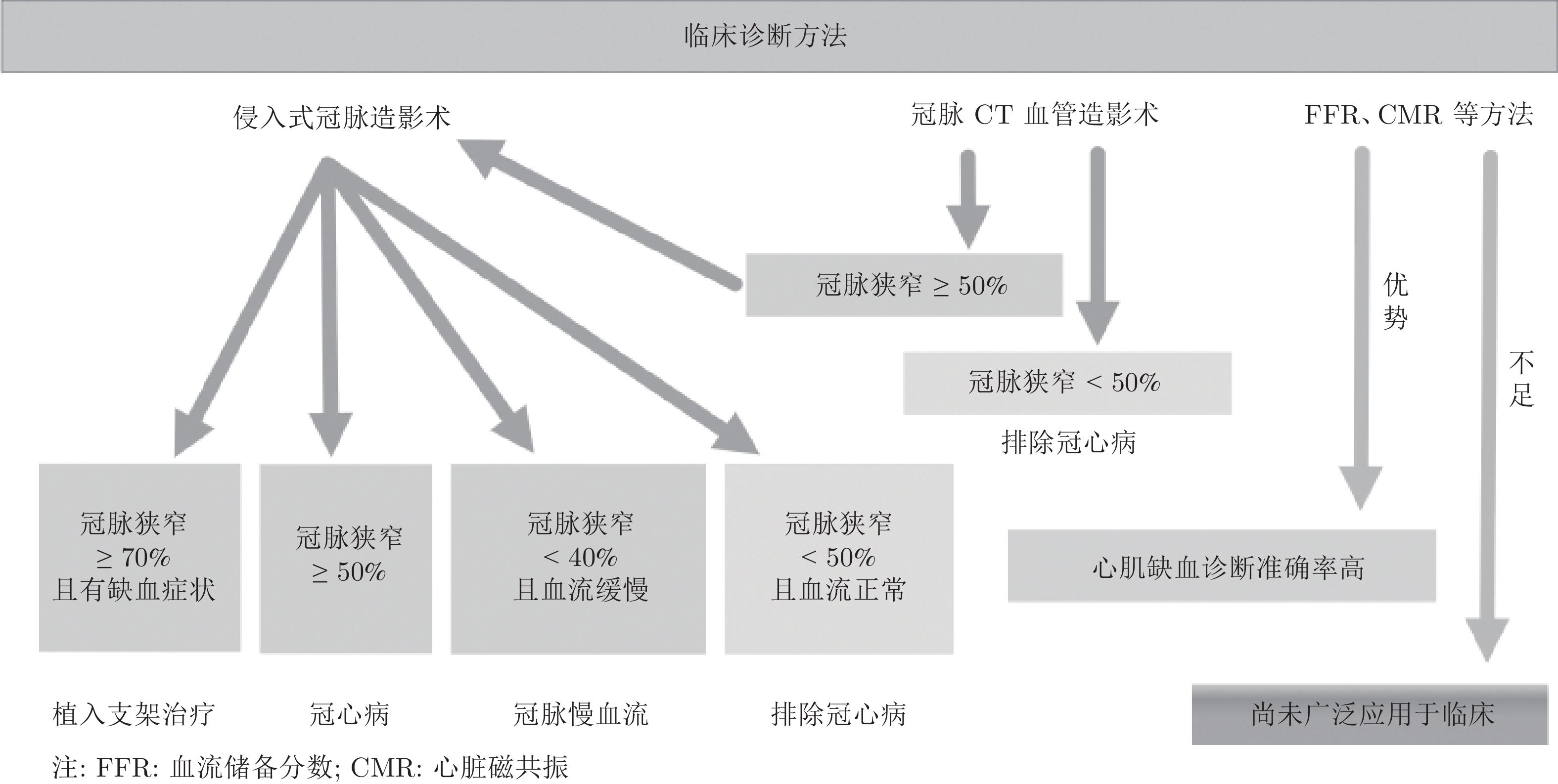
Early Detection of Myocardial Ischemia Based on Deterministic Learning and Cardiodynamicsgram
-
摘要: 心肌缺血早期检测是心血管疾病领域重要且困难的问题. 本文采用心电动力学图(Cardiodynamicsgram, CDG)开展心电图正常及大致正常时的心肌缺血早期检测研究. 1) 在分析已有基于心电图的心肌缺血检测方法所取得的进展及不足基础上, 构建一个既有心电图发生缺血性改变、又有心电图正常及大致正常、且包括经冠脉造影检验为冠脉阻塞性病变和非阻塞性病变的较大规模心肌缺血数据集. 2) 针对上述数据集中393例心电图正常及大致正常患者, 利用确定学习生成每份心电图的心电动力学图, 提取对心肌缺血和非缺血具有显著区分能力的心电动力学特征. 并以冠脉狭窄
$ \ge$ 50%为缺血标准, 采用机器学习算法构建心肌缺血检测模型. 3) 针对上述试验中假阳性病例, 利用由确定学习生成的具有明确物理意义的心电动力学图进行逐例分析, 发现其中许多假阳性存在慢血流现象(即冠脉非阻塞性病变). 对这些慢血流病例重新进行缺血标注, 以改善心肌缺血数据集标注精度. 通过上述三个步骤构建了更为准确的心肌缺血检测模型, 其缺血检测结果: 灵敏度90.1%、特异度85.2%、准确率89.0%和受试者工作特征曲线(Receiver operating characteristic curve, ROC)下面积(Area under curve, AUC) 0.93. 综上, 本文所构建的较大规模心肌缺血数据集可为心肌缺血检测研究和临床研究提供重要的数据基础; 且构建的心肌缺血检测模型对心电图正常及大致正常患者具有较强的缺血检测能力; 特别是, 由确定学习生成的心电动力学图具有较好的可解释性, 有助于发现缺血数据标注的偏差和模型的错误, 提高心肌缺血检测准确率.Abstract: Early detection of myocardial ischemia is a crucial and challenging problem in cardiovascular disease. In this paper, early detection of myocardial ischemia with normal or nearly normal electrocardiogram (ECG) is investigated by using cardiodynamicsgram (CDG). Firstly, by analyzing the advantages and disadvantages of existing ECG-based machine learning methods for myocardial ischemia detection, a relatively large-scale myocardial ischemia dataset is constructed, which contains ischemic, nearly normal and normal ECGs, as well as coronary stenosis and non-coronary stenosis detected by coronary angiography (CAG). Secondly, for 393 patients in the above dataset with normal or nearly normal ECGs, the deterministic learning algorithm is employed to generate CDGs, and the dynamical features underlying ECGs are extracted. The ischemia of patients is defined by coronary stenosis$ \ge$ 50%, and the detection model of myocardial ischemia is established using machine learning algorithms. It is shown that ischemia detection model can distinguish myocardial ischemia from non-ischemia effectively. Thirdly, by analyzing the false-positive cases in the above trial, in which each CDG is generated with clear physical meanings, coronary slow flow phenomenons (i.e., non-obstructive coronary lesions) are found in many of the false-positive cases. These cases are re-labeled as ischemia, and a more accurate model for ischemia detection is constructed, with the sensitivity of 90.1%, specificity of 85.2%, accuracy of 89.0% and AUC (area under curve) of 0.93, respectively. As such, in this paper a relatively large-scale myocardial ischemia dataset is constructed which will provide an essential basis for future research on detection algorithms and clinical trials of myocardial ischemia. The established model has the ability to detect ischemia from patients with normal or nearly normal ECGs. Particularly, the CDGs generated by deterministic learning have favorable interpretability, which is helpful for finding the deviations of ischemic data labeling and model errors, and is capable of improving the accuracy of myocardial ischemia detection. -
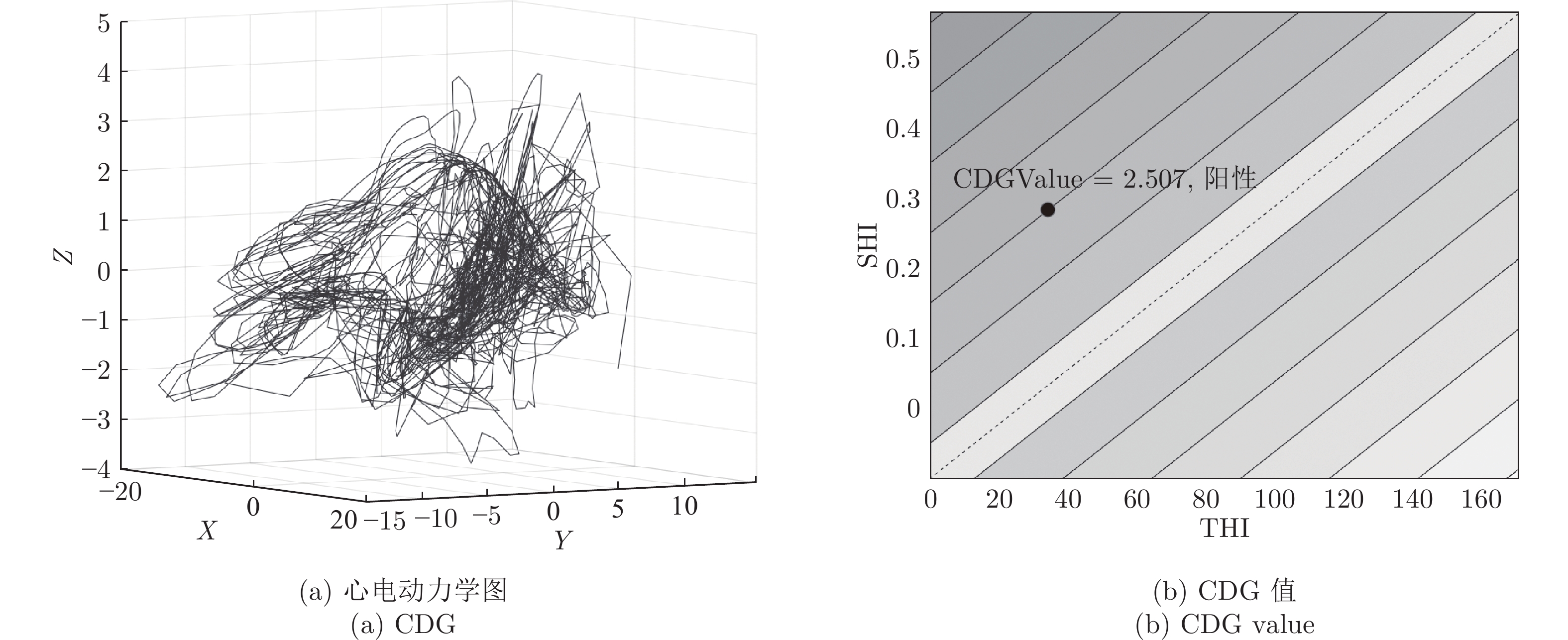
图 4 一例心肌缺血患者的心电动力学图及CDG值
Fig. 4 The CDG and CDG value of a patient with myocardial ischemia

图 5 冠脉狭窄与非狭窄组间的CDG值差异(
$ \lozenge $ :$ p<0.01 $ 存在差异有高度统计显著性;$ \bigstar $ : 超出边界的实例.)Fig. 5 Differences of CDG values between coronary stenosis and non-stenosis groups (
$ \lozenge $ :$ p<0.01 $ was considered as statistically significant.$ \bigstar $ : subjects that were out of boundaries.)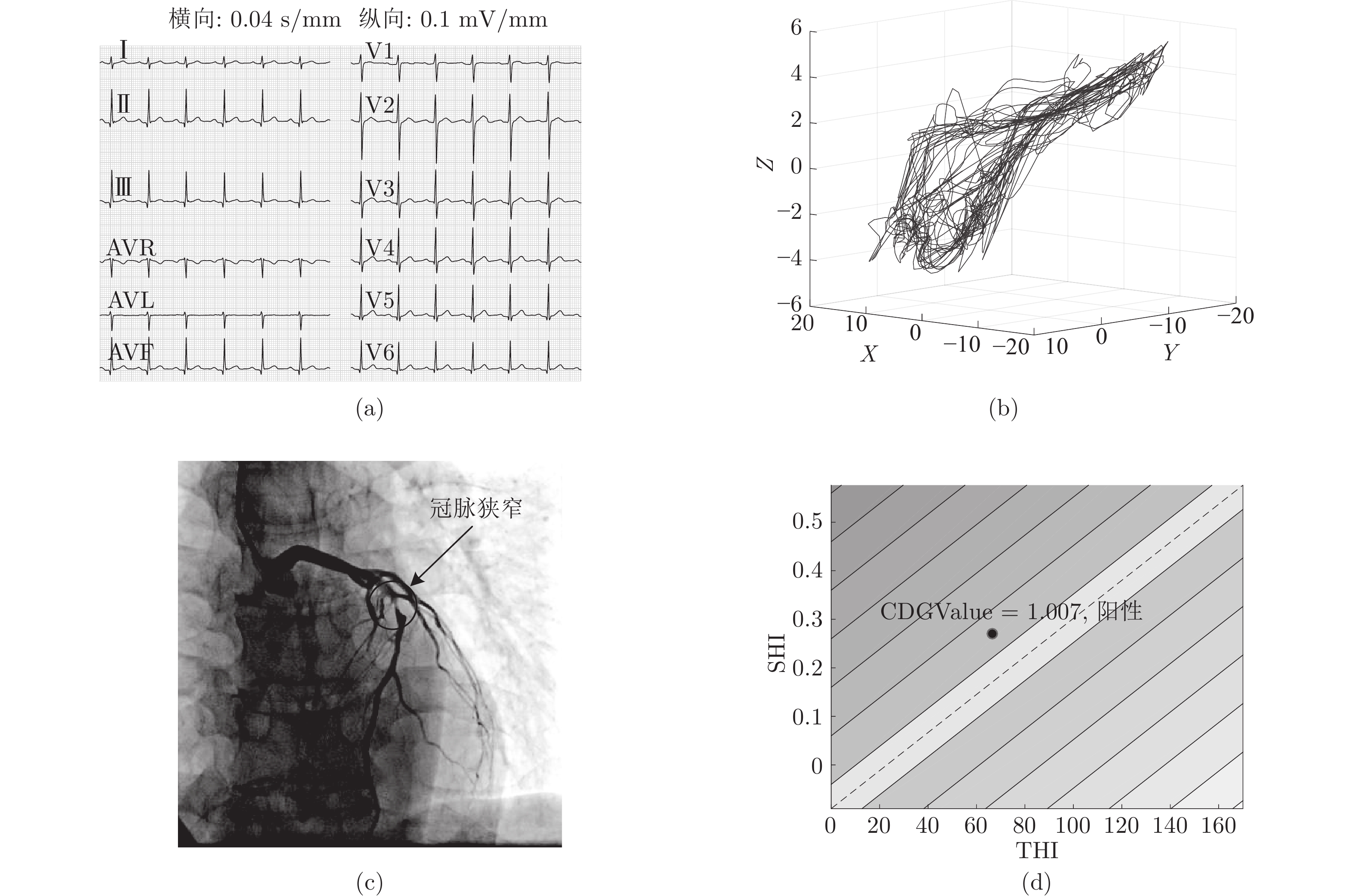
图 7 一例冠脉单支病变男性患者, 55岁 ((a) 正常心电图; (b) 心电动力学图散乱;(c) 冠脉前降支存在80%狭窄;(d) CDG值阳性)
Fig. 7 A case of ischemic male patient with single vessel disease, 55 years old ((a) Nondiagnostic ECG; (b) Irregular CDG; (c) The left anterior descending branch of the coronary artery is with stenosis 80%; (d) The positive CDG value)
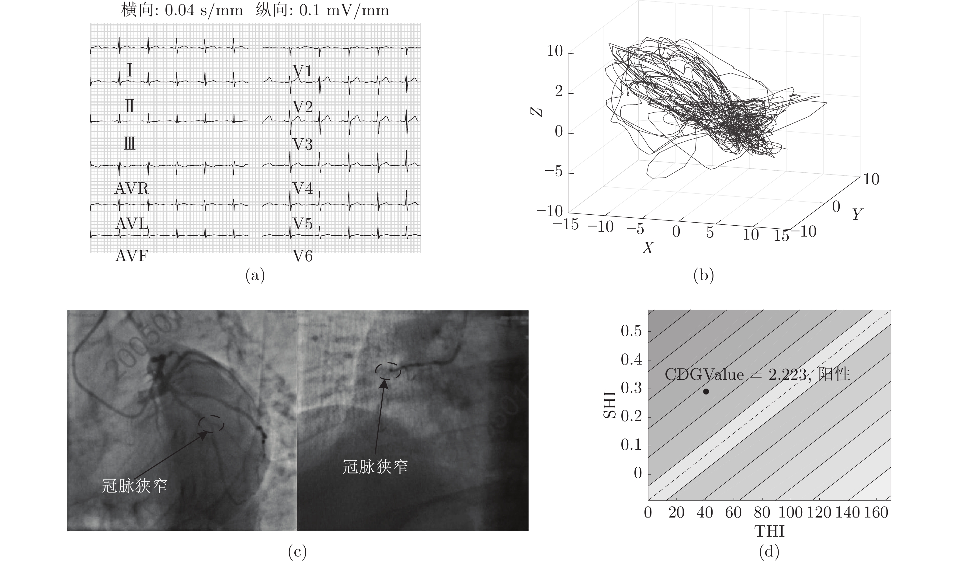
图 8 一例冠脉双支病变男性患者, 35岁 ((a) 正常心电图; (b) 心电动力学图散乱; (c) 冠脉回旋支中段50%狭窄,右冠近段100%狭窄; (d) CDG值阳性)
Fig. 8 A case of ischemic male patient with double-vessel disease, 35 years old ((a) Nondiagnostic ECG; (b) Irregular CDG; (c) The middle segment of the left circumflex artery is with stenosis 50% narrow, and the proximal segment of the right coronary artery is occluded; (d) The positive CDG value)

图 9 一例冠脉三支病变男性患者, 50岁 ((a) 正常心电图; (b) 心电动力学图散乱; (c) 中间支开口90%狭窄, 回旋支远段80%局限狭窄, 右冠远段90%局限狭窄; (d) CDG值阳性)
Fig. 9 A case of ischemic male patient with triple-vessel disease, 50 years old ((a) Nondiagnostic ECG; (b) Irregular CDG; (c) The opening of the middle branch is 90% narrow, the distal segment of the left circumflex artery is with stenosis 80%, and the distal segment of the right coronary artery is with stenosis 90%; (d) The positive CDG value)

图 10 一例非缺血女性患者, 47岁 ((a) 正常心电图; (b) 心电动力学图较为规整; (c) 正常冠脉; (d) CDG值阴性)
Fig. 10 A case of nonischemic female patient, 47 years old ((a) Normal ECG; (b) Regular CDG; (c) Normal coronary angiography; (d) The negative CDG value)
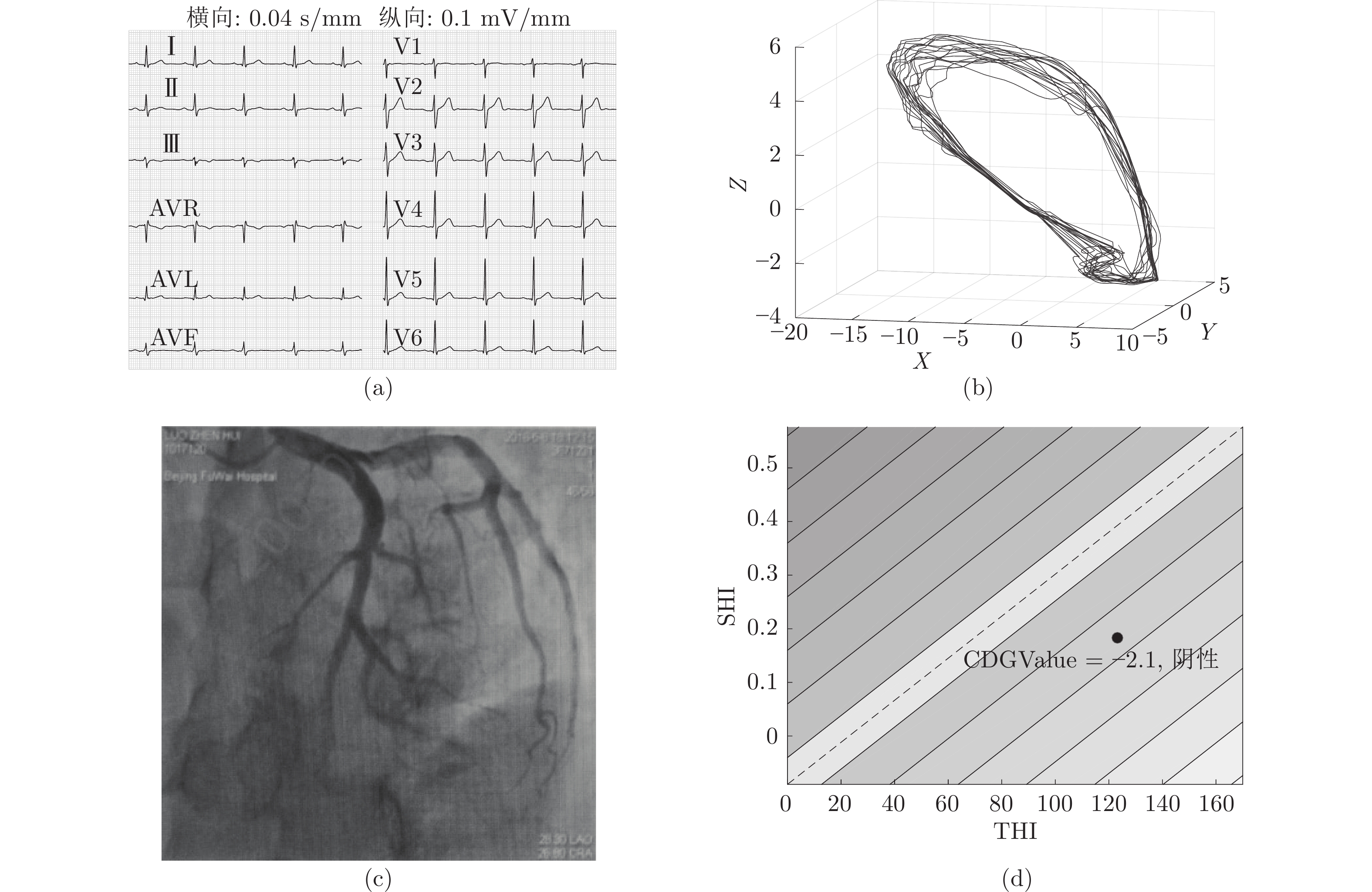
图 11 一例非缺血男性患者, 47岁 ((a) 正常心电图; (b) 心电动力学图规整; (c) 正常冠脉; (d) CDG值阴性)
Fig. 11 A case of nonischemic male patient, 47 years old ((a) Normal ECG; (b) Regular CDG; (c) Normal coronary angiography; (d) The negative CDG value)

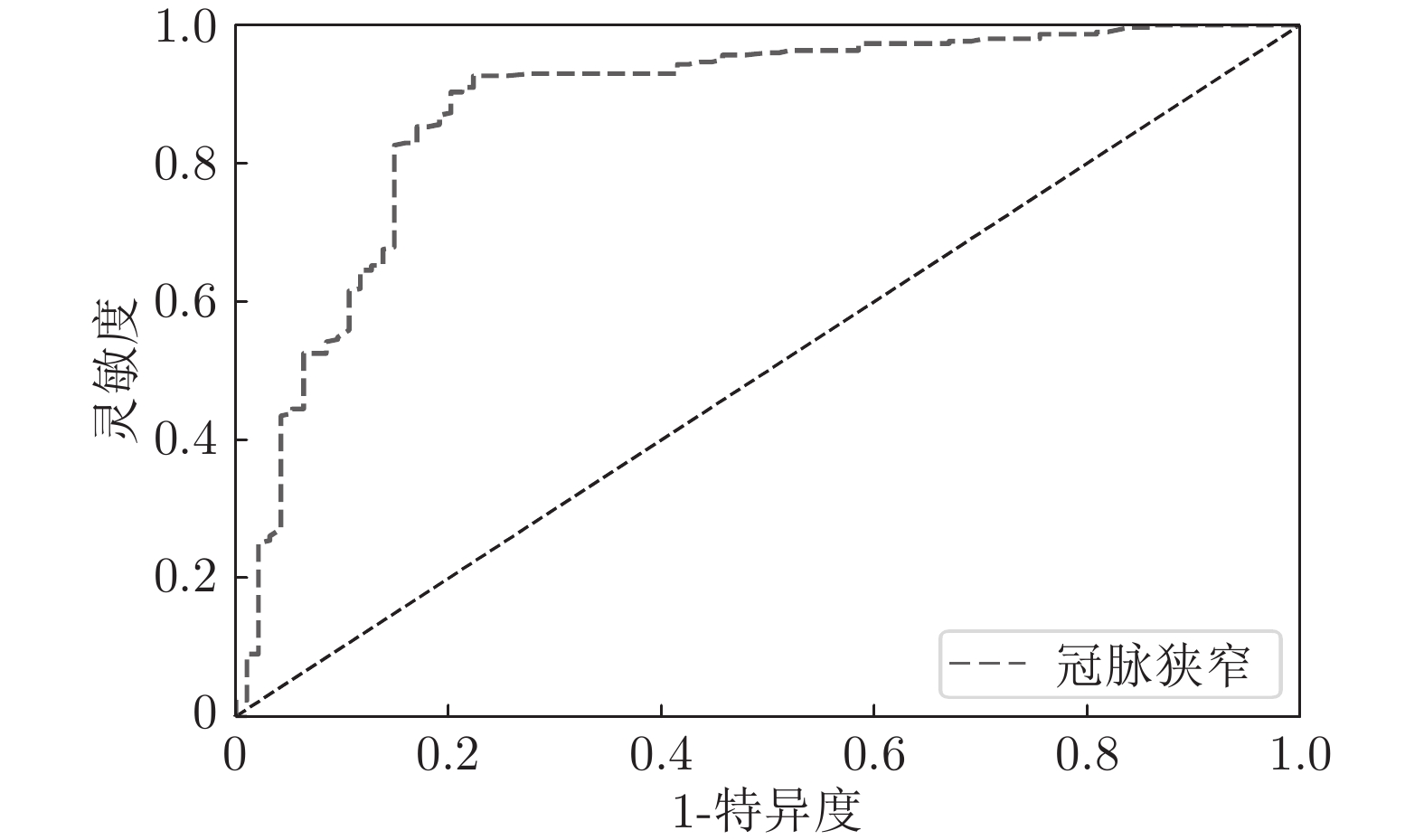
图 12 不同缺血标注精度下分类模型的ROC曲线
Fig. 12 ROC curves of classification models at different accuracy of ischemic labeling
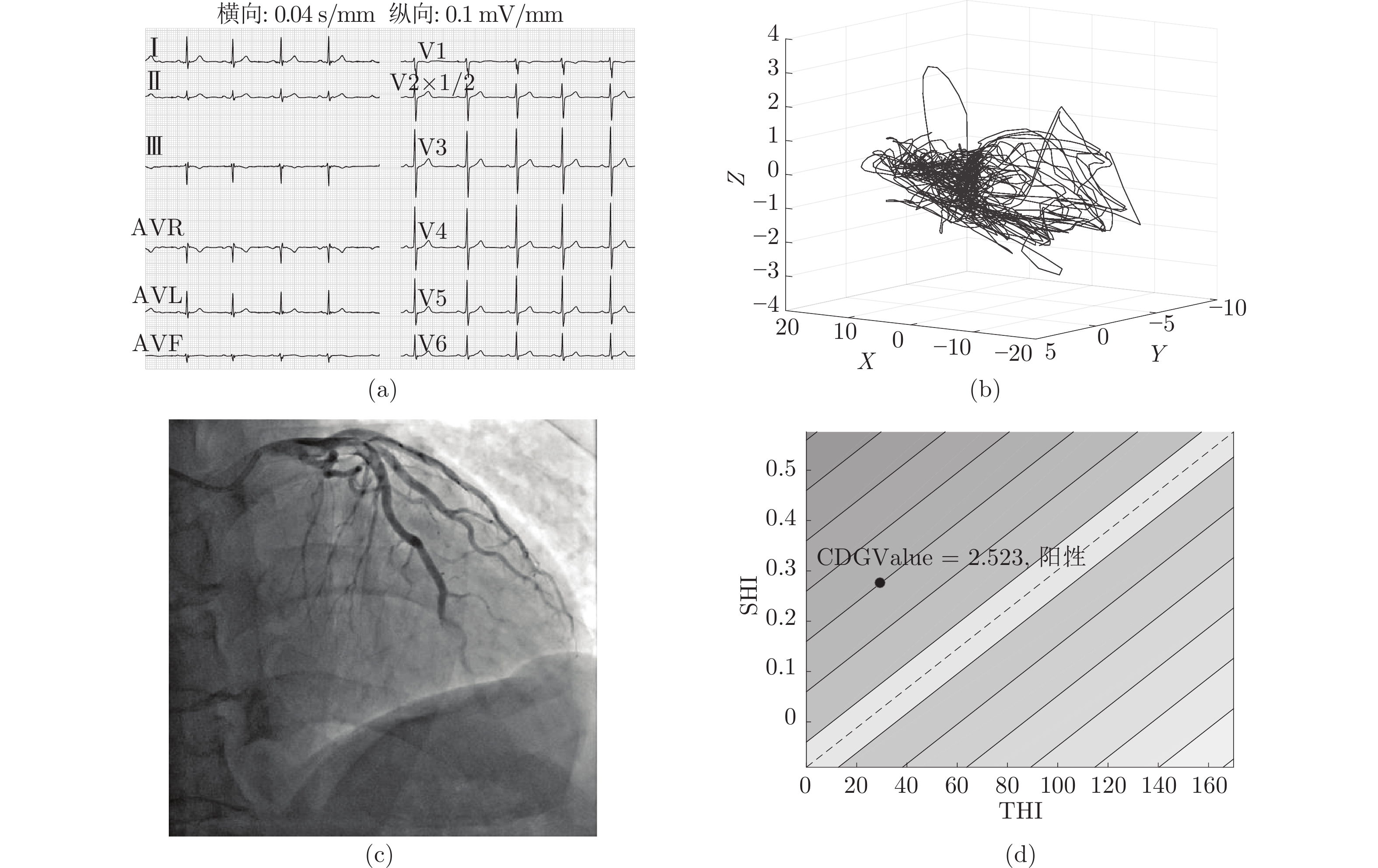
图 13 一例慢血流男性患者, 48岁 ((a) 正常心电图; (b) 心电动力学图散乱; (c) 冠脉无狭窄前降支慢血流; (d) CDG值阳性)
Fig. 13 A case of ischemic male patient with slow coronary flow, 48 years old ((a) Normal ECG; (b) Irregular CDG; (c) The left anterior descending branch of the coronary artery is with coronary slow flow; (d) The positive CDG value)
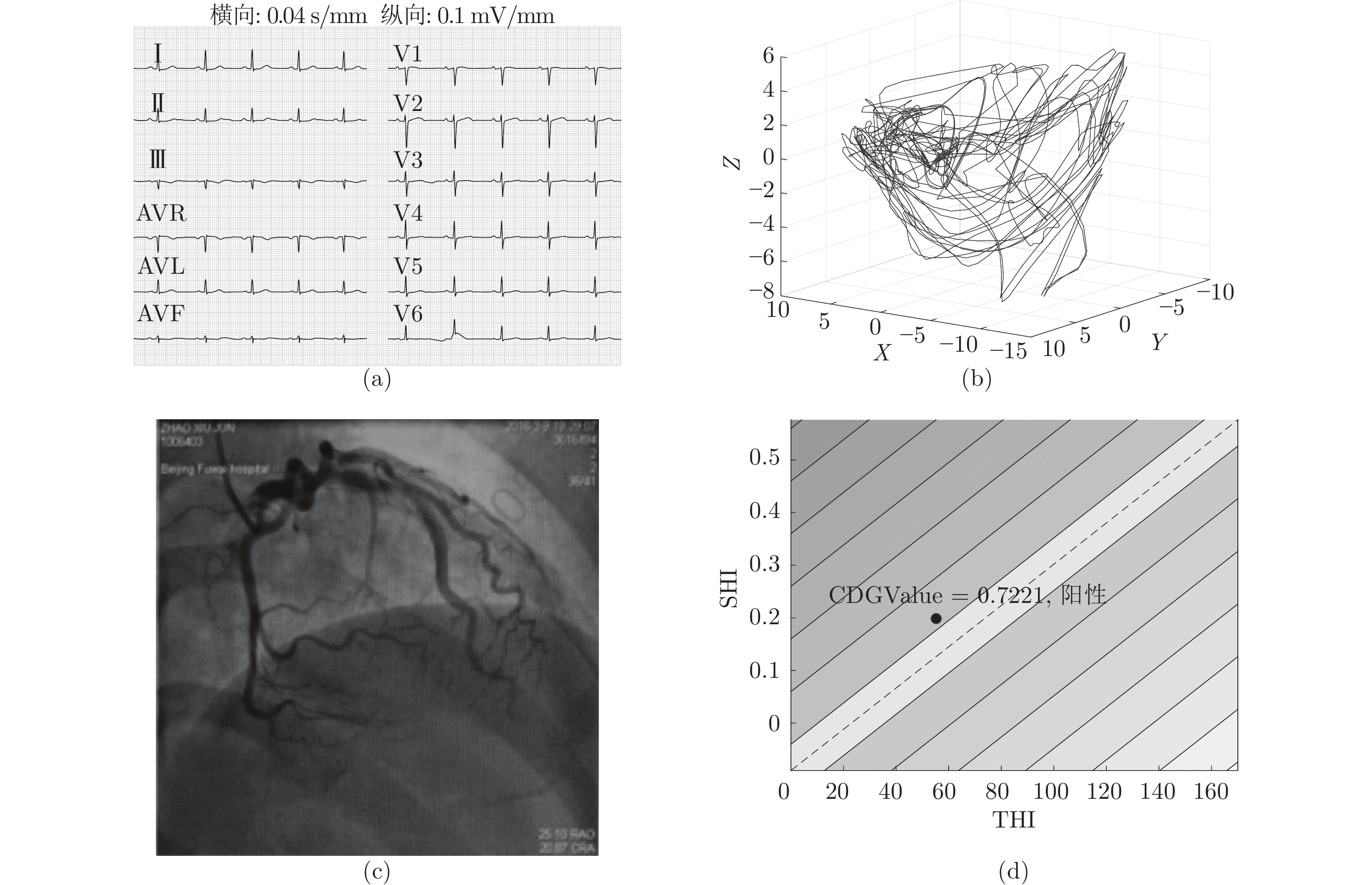
图 14 一例慢血流女性患者, 50岁 ((a) 正常心电图; (b) 心电动力学图散乱; (c) 冠脉无狭窄但前降支中段第一对角支慢血流; (d) CDG值阳性)
Fig. 14 A case of ischemic female patient with slow coronary flow, 50 years old ((a) Normal ECG; (b) Irregular CDG; (c) Coronary slow flow in the first diagonal branch of the coronary artery; (d) The positive CDG value)
表 1 疑似心肌缺血患者病例信息记录
Table 1 A case of suspected myocardial ischemic patient
项目 信息记录 编号/来源 SHZ2944/石河子市人民医院 年龄/性别 59/男 心率/血压 68 (次/分)/200 (高), 100 (低) (mmHg) 主诉 半月前无诱因再次出现胸骨中下段拳头大小范围压迫样疼痛, 伴胸闷、心慌、出汗, 症状持续数分钟休息后缓解, 症状频繁发作, 偶有静息下发作 既往史 平素健康状况一般, 高血压 30 年, 最高达 200/100 mmHg, 无其他病史 心电图 窦性心律, 偶发室早, T波改变 冠脉造影 前降支近段斑块; 回旋支近段斑块、远段 100% 闭塞, 可见前降支到回旋支侧枝形成; 右冠中段 80% 病变, 远段 90% 弥漫性病变 临床诊断 1) 冠心病, 不稳定性心绞痛; 2) 高血压 3 级 (很高危)  下载: 导出CSV
表 2 自建数据集与PTB数据集对比
Table 2 Comparison between PTB diagnostic dataset and the proposed dataset
来源 PTB 自建 总病例数 290 781 缺血病例 148 700 非缺血病例 52 81 心电图 基本发生缺血性改变 393 例正常或非特异性改变 缺血病因 冠脉狭窄 冠脉狭窄、慢血流
下载: 导出CSV
表 3 心电图正常或大致正常患者的人口基线特征
Table 3 Baseline characteristics of patients with normal or nearly normal ECG
类型 冠脉狭窄 (299) 非冠脉狭窄 (n = 94) p 值 冠脉慢血流 (13) 非冠脉病变 (81) 性别 (男性) 216/299 (72.2%) 9/13 (69.2%) 43/81 (53.7%) 0.005** 年龄 58±10 56±9 54±10 0.022* 收缩压 (mmHg) 129±10 131±18 127±14 0.563 舒张压 (mmHg) 77±10 85±14 77±10 0.109 心率 (beats/min) 72±10 65±6 71±10 0.012* 高血压 171/299 (57.2%) 10/13 (76.9%) 42/81 (51.9%) 0.226 糖尿病 88/299 (29.4%) 6/13 (46.2%) 18/81 (22.2%) 0.159 血脂异常 190/299 (63.5%) 8/13 (61.5%) 53/81 (65.4%) 0.937 注: 所有数据采用软件 SPSS 21.0 进行统计分析; 计量资料采用 Mann-Whitney 秩和检验, 表示为 (均值±标准差); 计数资料采用卡方检验, 用%表示; *: p < 0.05为差异有统计显著性; **: p < 0.01为差异有高度统计显著性.
下载: 导出CSV
表 4 不同缺血标注精度下, 心电动力学图的缺血检测结果
Table 4 The results of CDG in the detection of ischemia at different precision of ischemia labeling
缺血标准 灵敏度 (%) 特异度 (%) 准确率 (%) AUC 冠脉狭窄 85.1 82.6 87.8 0.88 冠脉狭窄及慢血流 90.1 85.2 89.0 0.93
下载: 导出CSV
表 5 本文方法与文献中的方法在PTB数据集上的心肌缺血检测结果对比
Table 5 Comparison of the CDG against the related literatures about myocardial ischemia detection
方法 数据 方法特点 特征数 分类器 性能 (%) 准确率 敏感度 特异度 Sharma等 (2015)[20] 导联: 12 导联心电记录: 148 MI, 52 HC 多尺度小波能量特征 72 KNN/SVM 96.00 93.00 99.00 Han等 (2019)[15] 导联: 12 导联心电记录: 28 213 MI,
5 373 HC能量熵; 形态学特征 22 SVM 92.69 80.96 80.96 Diker等 (2018[17] 导联: 不可知心电信号: 148 MI, 52 HC 形态学特征; 时域特征;
离散小波变换特征9 SVM 87.80 86.97 88.67 Sharma等 (2018)[18] 导联: II、III、aVF 导联心电信号: 3 240
下壁 MI, 3 037 HC样本熵; 归一化子带能量;
对数能量熵; 中值斜率10 KNN/SVM 81.71 79.01 79.26 Acharya等 (2017)[27] 导联: II 导联心拍: 40 182 MI, 10 546 HC 卷积神经网络 − 全连接网络 95.22 95.49 94.19 Han等 (2020)[29] 导联: 12 导联心电记录: 17 212 MI,
6 945 HC多导联残差网络 − 全连接 softmax 95.49 94.85 97.37 本文方法 导联: 12 导联心电记录: 148 MI, 52 HC 心电动力学图特征 2 SVM-Linear 97.00 98.65 92.31
下载: 导出CSV
-
[1] World Health Organization. The top 10 causes of death [Online], available: https://www.who.int/en/news-room/fact-sheets/detail/the-top-10-causes-of-death, June 16, 2020 [2] 国家卫生计生委合理用药专家委员会, 中国药师协会. 冠心病合理用药指南(第2版). 中国医学前沿杂志(电子版), 2018, 10(6): 1−130Committee of Experts on Rational Drug Use National Health and Family Planning Commission of the P. R. China, Chinese Pharmacists Association. Guideline on rational use of drugs for coronary heart disease (2nd edition). Chinese Journal of the Frontiers of Medical Science (Electronic Version), 2018, 10(6): 1−130 [3] Tamis-Holland J E, Jneid H, Reynolds H R, Agewall S, Brilakis E S, Brown T M, et al. Contemporary diagnosis and management of patients with myocardial infarction in the absence of obstructive coronary artery disease: A scientific statement from the American Heart Association. Circulation, 2019, 139(18): e891−e908 [4] Song Y B, Arbab-Zadeh A, Matheson M B, Ostovaneh M R, Vavere A, Dewey M, et al. Contemporary discrepancies of stenosis assessment by computed tomography and invasive coronary angiography. Circulation: Cardiovascular Imaging, 2019, 12(2): e007720 [5] 中华医学会心血管病学分会介入心脏病学组, 中华医学会心血管病学分会动脉粥样硬化与冠心病学组, 中国医师协会心血管内科医师分会血栓防治专业委员会, 中华心血管病杂志编辑委员会. 稳定性冠心病诊断与治疗指南. 中华心血管病杂志, 2018, 46(9): 680−694 doi: 10.3760/cma.j.issn.0253-3758.2018.09.004Interventional Cardiology Group in Chinese Society of Cardiology, Atherosclerosis and Coronary Artery Disease Group in Chinese Society of Cardiology, Chinese College of Cardiovascular Physicians, Editorial Board of Chinese Journal of Cardiology. Guideline for the diagnosis and treatment of stable coronary artery disease. Chinese Journal of Cardiology, 2018, 46(9): 680−694 doi: 10.3760/cma.j.issn.0253-3758.2018.09.004 [6] Colombo A, Panoulas V F. Diagnostic coronary angiography is getting old!. JACC: Cardiovascular Imaging, 2015, 8(1): 11−13 doi: 10.1016/j.jcmg.2014.11.003 [7] Tonino P A L, Fearon W F, De Bruyne B, Oldroyd K G, Leesar M A, Ver Lee P N, et al. Angiographic versus functional severity of coronary artery stenoses in the FAME study: Fractional flow reserve versus angiography in multivessel evaluation. Journal of the American College of Cardiology, 2010, 55(25): 2816−2821 doi: 10.1016/j.jacc.2009.11.096 [8] Park H B, Heo R, óHartaigh B, Cho I, Gransar H, Nakazato R, et al. Atherosclerotic plaque characteristics by CT angiography identify coronary lesions that cause ischemia: A direct comparison to fractional flow reserve. JACC: Cardiovascular Imaging, 2015, 8(1): 1−10 doi: 10.1016/j.jcmg.2014.11.002 [9] 王建安. 透过现象看本质: 从冠状动脉狭窄与心肌缺血的辩证关系说起. 中华心血管病杂志, 2018, 46(9): 671 doi: 10.3760/cma.j.issn.0253-3758.2018.09.001Wang Jian-An. Essence through the appearance: The relationship between coronary artery stenosis and myocardial ischemia is discussed. Chinese Journal of Cardiology, 2018, 46(9): 671 doi: 10.3760/cma.j.issn.0253-3758.2018.09.001 [10] 中华医学会心血管病学分会基础研究学组, 中华医学会心血管病学分会介入心脏病学组, 中华医学会心血管病学分会女性心脏健康学组, 中华医学会心血管病学分会动脉粥样硬化和冠心病学组. 冠状动脉微血管疾病诊断和治疗的中国专家共识. 中国循环杂志, 2017, 32(5): 421−430 doi: 10.3969/j.issn.1000-3614.2017.05.003Basic Research Group, Interventional Cardiology Group, Female Heart Health Greup, and Atherosclerosis and Coronary Heart Disease Group of Chinese Society of Cardiology. Recommendation of Chinese experts on the diagnosis and treatment of coronary microvascular disease. Chinese Circulation Journal, 2017, 32(5): 421−430 doi: 10.3969/j.issn.1000-3614.2017.05.003 [11] Thygesen K, Alpert J S, Jaffe A S, Chaitman B R, Bax J J, Morrow D A, et al. Fourth universal definition of myocardial infarction (2018). European Heart Journal, 2019, 40(3): 237−269 doi: 10.1093/eurheartj/ehy462 [12] Lilly L S. Pathophysiology of Heart Disease: A Collaborative Project of Medical Students and Faculty (July edition). New York: Wolters Kluwer, 2020. 134−153 [13] Drew B J, Pelter M M, Lee E, Zegre J, Schindler D, Fleischmann K E. Designing prehospital ECG systems for acute coronary syndromes. Lessons learned from clinical trials involving 12-lead ST-segment monitoring. Journal of Electrocardiology, 2005, 38(4 Suppl): 180−185 [14] Rouan G W, Lee T H, Cook E F, Brand D A, Weisberg M C, Goldman L. Clinical characteristics and outcome of acute myocardial infarction in patients with initially normal or nonspecific electrocardiograms (a report from the Multicenter Chest Pain Study). The American Journal of Cardiology, 1989, 64(18): 1087−1092 doi: 10.1016/0002-9149(89)90857-6 [15] Han C, Shi L. Automated interpretable detection of myocardial infarction fusing energy entropy and morphological features. Computer Methods and Programs in Biomedicine, 2019, 175: 9−23 doi: 10.1016/j.cmpb.2019.03.012 [16] Sadhukhan D, Pal S, Mitra M. Automated identification of myocardial infarction using harmonic phase distribution pattern of ECG data. IEEE Transactions on Instrumentation and Measurement, 2018, 67(10): 2303−2313 doi: 10.1109/TIM.2018.2816458 [17] Diker A, Cömert Z, Avci E, Velappan S. Intelligent system based on Genetic Algorithm and support vector machine for detection of myocardial infarction from ECG signals. In: Proceedings of the 26th Signal Processing and Communications Applications Conference (SIU). Izmir, Turkey: IEEE, 2018. 1−4 [18] Sharma L D, Sunkaria R K. Inferior myocardial infarction detection using stationary wavelet transform and machine learning approach. Signal, Image and Video Processing, 2018, 12(2): 199−206 doi: 10.1007/s11760-017-1146-z [19] Papaloukas C, Fotiadis D I, Likas A, Michalis L K. An ischemia detection method based on artificial neural networks. Artificial Intelligence in Medicine, 2002, 24(2): 167−178 doi: 10.1016/S0933-3657(01)00100-2 [20] Sharma L N, Tripathy R K, Dandapat S. Multiscale energy and eigenspace approach to detection and localization of myocardial infarction. IEEE Transactions on Biomedical Engineering, 2015, 62(7): 1827−1837 doi: 10.1109/TBME.2015.2405134 [21] Ansari S, Farzaneh N, Duda M, Horan K, Andersson H B, Goldberger Z D, et al. A review of automated methods for detection of myocardial ischemia and infarction using electrocardiogram and electronic health records. IEEE Reviews in Biomedical Engineering, 2017, 10: 264−298 doi: 10.1109/RBME.2017.2757953 [22] Goletsis Y, Papaloukas C, Fotiadis D I, Likas A, Michalis L K. Automated ischemic beat classification using genetic algorithms and multicriteria decision analysis. IEEE Transactions on Biomedical Engineering, 2004, 51(10): 1717−1725 doi: 10.1109/TBME.2004.828033 [23] Dohare A K, Kumar V, Kumar R. Detection of myocardial infarction in 12 lead ECG using support vector machine. Applied Soft Computing, 2018, 64: 138−147 doi: 10.1016/j.asoc.2017.12.001 [24] Acharya U R, Fujita H, Adam M, Lih O S, Sudarshan V K, Hong T J, et al. Automated characterization and classification of coronary artery disease and myocardial infarction by decomposition of ECG signals: A comparative study. Information Sciences, 2017, 377: 17−29 doi: 10.1016/j.ins.2016.10.013 [25] Bousseljot R, Kreiseler D, Schnabel A. Nutzung der EKG-signaldatenbank cardiodat der PTB über das internet. Biomedizinische Technik, 1995, 40(1): 317−318 [26] Taddei A, Distante G, Emdin M, Pisani P, Moody G B, Zeelenberg C, et al. The European ST-T database: Standard for evaluating systems for the analysis of ST-T changes in ambulatory electrocardiography. European Heart Journal, 1992, 13(9): 1164−1172 doi: 10.1093/oxfordjournals.eurheartj.a060332 [27] Acharya U R, Fujita H, Oh S L, Hagiwara Y, Tan J H, Adam M. Application of deep convolutional neural network for automated detection of myocardial infarction using ECG signals. Information Sciences, 2017, 415-416: 190−198 doi: 10.1016/j.ins.2017.06.027 [28] Tan J H, Hagiwara Y, Pang W, Lim I, Oh S L, Adam M, et al. Application of stacked convolutional and long short-term memory network for accurate identification of CAD ECG signals. Computers in Biology and Medicine, 2018, 94: 19−26 doi: 10.1016/j.compbiomed.2017.12.023 [29] Han C, Shi L. ML–ResNet: A novel network to detect and locate myocardial infarction using 12 leads ECG. Computer Methods and Programs in Biomedicine, 2020, 185: Article No. 105138 [30] Rajkomar A, Dean J, Kohane I. Machine learning in medicine. New England Journal of Medicine, 2019, 380(14): 1347−1358 doi: 10.1056/NEJMra1814259 [31] Wang C, Dong X D, Ou S X, Wang W, Hu J M, Yang F F. A new method for early detection of myocardial ischemia: Cardiodynamicsgram (CDG). Science China Information Sciences, 2016, 59(1): 1−11 [32] Wang C, Hill D J. Deterministic Learning Theory for Identification, Recognition, and Control. Boca Raton: CRC Press, 2009. [33] Deng M Q, Tang M, Wang C, Shan L, Zhang L F, Zhang J T, et al. Cardiodynamicsgram as a new diagnostic tool in coronary artery disease patients with nondiagnostic electrocardiograms. The American Journal of Cardiology, 2017, 119(5): 698−704 doi: 10.1016/j.amjcard.2016.11.028 [34] Jurko S, Rozinaj G. High resolution of the ECG signal by polynomial approximation. Radioengineering, 2006, 15(1): 32−37 [35] 中华医学会心血管病学分会, 中华心血管病杂志编辑委员会. 推荐在我国采用心肌梗死全球统一定义. 中华心血管病杂志, 2008, 36(10): 867−869 doi: 10.3321/j.issn:0253-3758.2008.10.002Chinese Society of Cardiology, Editorial Board of Chinese Journal of Cardiology. Recommendation of the application of universal definition of myocardial infarction in China. Chinese Journal of Cardiology, 2008, 36(10): 867−869 doi: 10.3321/j.issn:0253-3758.2008.10.002 [36] 何永福. 冠状动脉慢血流现象病理生理机制及治疗进展. 心血管病学进展, 2018, 39(3): 448−452He Yong-Fu. Pathophysiological mechanism and treatment of slow blood flow in coronary artery. Advances in Cardiovascular Diseases, 2018, 39(3): 448−452 [37] 王帅, 薛强, 刘毅, 关欣, 陶凌. 大样本冠状动脉慢血流的相关因素分析. 心脏杂志, 2017, 29(2): 180−183Wang Shuai, Xue Qiang, Liu Yi, Guan Xin, Tao Ling. Analysis of risk factors for slow coronary flow in 958 patients. Chinese Heart Journal, 2017, 29(2): 180−183 [38] Hawkins B M, Stavrakis S, Rousan T A, Abu-Fadel M, Schechter E. Coronary slow flow-prevalence and clinical correlations. Circulation Journal, 2012, 76(4): 936−942 doi: 10.1253/circj.CJ-11-0959 [39] Wang C, Hill D J. Deterministic learning and rapid dynamical pattern recognition. IEEE Transactions on Neural Networks, 2007, 18(3): 617−630 doi: 10.1109/TNN.2006.889496 [40] Wang C, Hill D J. Learning from neural control. IEEE Transactions on Neural Networks, 2006, 17(1): 130−146 doi: 10.1109/TNN.2005.860843 [41] Yuan C Z, Wang C. Design and performance analysis of deterministic learning of sampled-data nonlinear systems. Science China Information Sciences, 2014, 57(3): 1−18 [42] 王聪, 陈填锐, 刘腾飞. 确定学习与基于数据的建模及控制. 自动化学报, 2009, 35(6): 693−706 doi: 10.3724/SP.J.1004.2009.00693Wang Cong, Chen Tian-Rui, Liu Teng-Fei. Deterministic learning and data-based modeling and control. Acta Automatica Sinica, 2009, 35(6): 693−706 doi: 10.3724/SP.J.1004.2009.00693 [43] Wang C, Chen T R. Rapid detection of small oscillation faults via deterministic learning. IEEE Transactions on Neural Networks, 2011, 22(8): 1284−1296 doi: 10.1109/TNN.2011.2159622 [44] 王聪, 文彬鹤, 司文杰, 彭滔, 袁承志, 陈填锐等. 轴流压气机旋转失速建模与检测: I—基于确定学习理论与高阶Moore-Greitzer模型的研究. 自动化学报, 2014, 40(7): 1265−1277Wang Cong, Wen Bin-He, Si Wen-Jie, Peng Tao, Yuan Cheng-Zhi, Chen Tian-Rui, et al. Modeling and detection of rotating stall in axial flow compressors: Part I - Investigation on high-order M-G models via deterministic learning. Acta Automatica Sinica, 2014, 40(7): 1265−1277 [45] 王乾, 王聪. 基于确定学习理论和Lempel-Ziv复杂度的非线性系统动态特征提取. 自动化学报, 2018, 44(10): 1812−1823Wang Qian, Wang Cong. Dynamic feature extraction of nonlinear systems with deterministic learning theory and spatio-temporal Lempel-Ziv complexity. Acta Automatica Sinica, 2018, 44(10): 1812−1823 [46] Kors J A, Van Herpen G, Sittig A C, Van Bemmel J H. Reconstruction of the Frank vectorcardiogram from standard electrocardiographic leads: Diagnostic comparison of different methods. European Heart Journal, 1990, 11(12): 1083−1092 doi: 10.1093/oxfordjournals.eurheartj.a059647 [47] Dower G E, Machado H B. XYZ data interpreted by a 12-lead computer program using the derived electrocardiogram. Journal of Electrocardiology, 1979, 12(3): 249−261 doi: 10.1016/S0022-0736(79)80058-8 [48] Dower G E, Machado H B, Osborne J A. On deriving the electrocardiogram from vectorcardiographic leads. Clinical Cardiology, 1980, 3(2): 87−95 doi: 10.1002/clc.1980.3.2.87 [49] Dower G E, Yakush A, Nazzal S B, Jutzy R V, Ruiz C E. Deriving the 12-lead electrocardiogram from four (EASI) electrodes. Journal of Electrocardiology, 1988, 21 Suppl: S182−S187 [50] Zhang Z Z, Chen P J, McGough M, Xing F Y, Wang C B, Bui M, et al. Pathologist-level interpretable whole-slide cancer diagnosis with deep learning. Nature Machine Intelligence, 2019, 1(5): 236−245 doi: 10.1038/s42256-019-0052-1 [51] Guidotti R, Monreale A, Ruggieri S, Turini F, Giannotti F, Pedreschi D. A survey of methods for explaining black box models. ACM Computing Surveys, 2019, 51(5): Article No. 93 [52] 纪守领, 李进锋, 杜天宇, 李博. 机器学习模型可解释性方法、应用与安全研究综述. 计算机研究与发展, 2019, 56(10): 2071−2096 doi: 10.7544/issn1000-1239.2019.20190540Ji Shou-Ling, Li Jin-Feng, Du Tian-Yu, Li Bo. Survey on techniques, applications and security of machine learning interpretability. Journal of Computer Research and Development, 2019, 56(10): 2071−2096 doi: 10.7544/issn1000-1239.2019.20190540 [53] Verrier R L, Klingenheben T, Malik M, El-Sherif N, Exne D V, Hohnloser S H, et al. Microvolt T-wave alternans Physiological basis, methods of measurement, and clinical utility-consensus guideline by International Society for Holter and Noninvasive Electrocardiology. Journal of the American College of Cardiology, 2011, 58(13): 1309−1324 doi: 10.1016/j.jacc.2011.06.029 -


 下载:
下载:


计量
- 文章访问数: 2533
- HTML全文浏览量: 727
- PDF下载量: 241
- 被引次数: 0
