

-
摘要:
深度学习可以有效提取图像隐含特征,在医学影像识别方面的应用快速发展. 由于糖尿病视网膜病变(Diabetic retinopathy, DR)诊断标准明确、分类体系成熟,应用深度学习诊断糖尿病视网膜病变近年来成为研究热点. 本文从深度学习方法在DR诊断中的最新研究进展、DR诊断的一般流程、公共数据集、医学影像标注方法、主要实现模型、面临的主要挑战几方面, 对深度学习方法在糖尿病视网膜病变诊断中的应用进行了详细综述, 便于更多机器视觉、尤其是深度学习医学影像的研究者们参照对比,加快该领域研究的成熟度和临床落地应用.
Abstract:Deep learning can effectively extract the hidden features of image and its application in medical image recognition is developing rapidly. Due to the clear diagnostic criteria for diabetic retinopathy (DR) and the mature classification system, the application of deep learning to diagnose diabetic retinopathy has become a research hotspot in recent years. Therefore, this paper reviews the application of deep learning methods in the diagnosis of diabetic retinopathy detailedly based on the latest research progress of deep learning for DR diagnosis, the general flow for DR diagnosis, public dataset, medical image annotation method, main models and major challenges. It brings convenience for more researchers of computer vision deepling learning, especially medical imaging deep learning, to speed up the research maturity and clinical application in this field.
-
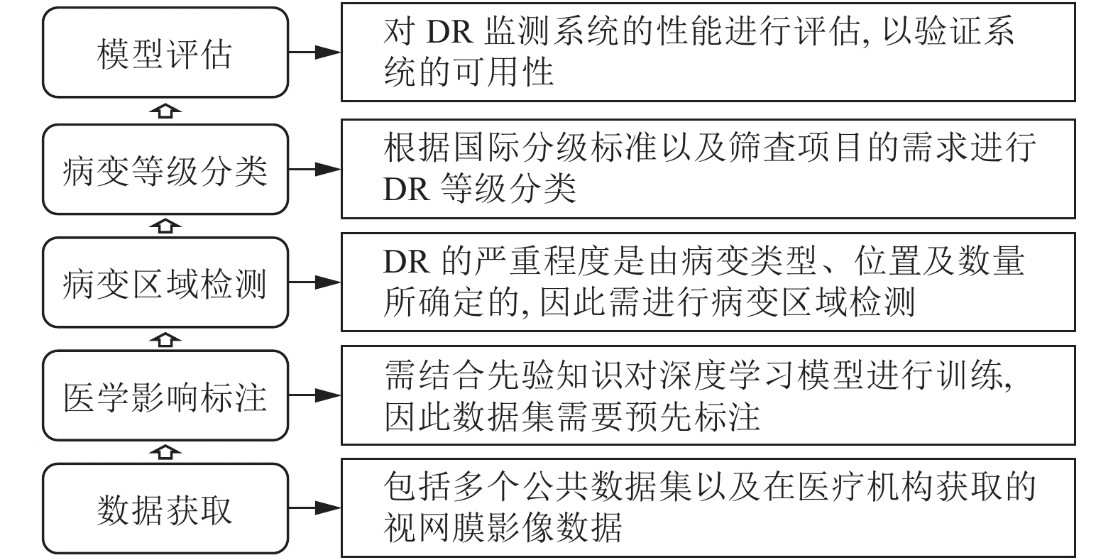
图 1 基于深度学习DR诊断的一般框架
Fig. 1 General framework of diabetic retinal diagnosis based on deep learning
表 1 糖尿病视网膜病变国际分级标准
Table 1 International classification of diabetic retinopathy and diabetic macular edema
糖尿病视网膜病变 散瞳后眼镜所见 随访建议 无视网膜病变 (无DR) 无异常 1 ~ 2 年随访一次 轻度非增殖性糖尿病视网膜
病变 (轻度NPDR)仅有微动脉瘤 1 ~ 2 年随访一次 中度非增殖性糖尿病视网膜
病变 (中度NPDR)比仅有微动脉瘤重, 比重者轻 半年到一年随访一次或转
诊至眼科医师重度非增殖性糖尿病视网
膜病变 (重度NPDR)有以下任一症状之一:
4个象限每个都有20个以上的内出血病灶;
2个以上象限有确定的静脉珠状改变;
1个以上象限有明显的视网膜
内微血管异常并且无增殖性病变体征转诊至眼科医师 增殖性糖尿病视网膜
病变 (PDR)具有重度非增殖性症状且有以下一种或多种情形:
新生血管, 玻璃体积血/视网膜前出血转诊至眼科医师  下载: 导出CSV
表 2 糖尿病视网膜病变公共数据集
Table 2 Public data set on diabetic retinal
名称 数据类型 图像信息 图片尺寸 图片格式 数据量 (幅) 获取权限 链接 IDRID[24] CFP 图像按照国际标准进行糖网和黄斑水肿分级, 并对其中 81 幅有糖网征象的图像进行了病变的像素级标注. 4228 × 2848 JPEG 516 注册 IEEE
账号获取https://ieee-dataport.org/open-access/indian-diabetic-retinopathy-image-dataset-idrid E-Ophtha[25] CFP 由 e-ophtha-MA (微动脉瘤) 和E-Ophtha-EX (渗出) 两个子数据库组成. 标注了 EX 和 MA 的区域, 以掩模的方式给出. 2544 × 1696
1440 × 960
1504 × 1000 等JPEG 463 提交邮箱以
获取下载码https://ieee-dataport.org/open-access/indian-diabetic-retinopathy-image-dataset-idrid DRiDB[26] CFP 每幅图像由至少5名专家标注, 标注内容包含所有视网膜主要解剖结构和病理特征以及糖网分级. 720 × 676 BMP 50 发送邮件来请求
访问数据库https://ipg.fer.hr/ipg/resources/image_database Messidor[27] CFP 图像标注了糖尿病性视网膜病变分级及黄斑水肿分级. 1400 × 960
2 240 × 1488
2304 × 1536TIFF 1200 提交邮箱以
获取下载码http://www.adcis.net/en/Download-Third-Party/Messidor.html Messidor-2[28] CFP 每幅图像标注了糖尿病性视网膜病变分级及黄斑水肿分级. 1400 × 960
2240 × 1488
2304 × 1536TIFF 1784 提交邮箱以
获取下载码http://latim.univ-brest.fr/indexfce0.html DIARETDB0[29] CFP 该数据集是用于糖尿病视网膜病变检测的基准的公开数据集, 每幅图像标注了病变信息. 1500 × 1152 PNG 130 直接下载 http://www.it.lut.fi/project/imageret/diaretdb0/index.html DIARETDB1[30] CFP 该数据集可用做基准糖尿病视网膜病变检测数据集, 由 4 名医学专家对糖网相关病变区域进行
标注.1500 × 1152 PNG 89 直接下载 http://www.it.lut.fi/project/imageret/diaretdb1/ Large dataset of OCT on Mendeley[15] OCT 图像分为训练集和测试集, 每个集合含有 4 种标签的数据: CNV, DME, DR-USEN 和 NORMAL. 不统一约为
400 × 500JPEG > 50000 直接下载 https://data.mendeley.com/archiver/rscbjbr9sj?version=3 Dataset for OCT classification[31] OCT 由 50 个正常、48 个干性 AMD 和 50 个 DME 组成. 765 × 765 TIFF 3700 直接下载 https://sites.google.com/site/hosseinrabbanikhorasgani/datasets-1
下载: 导出CSV
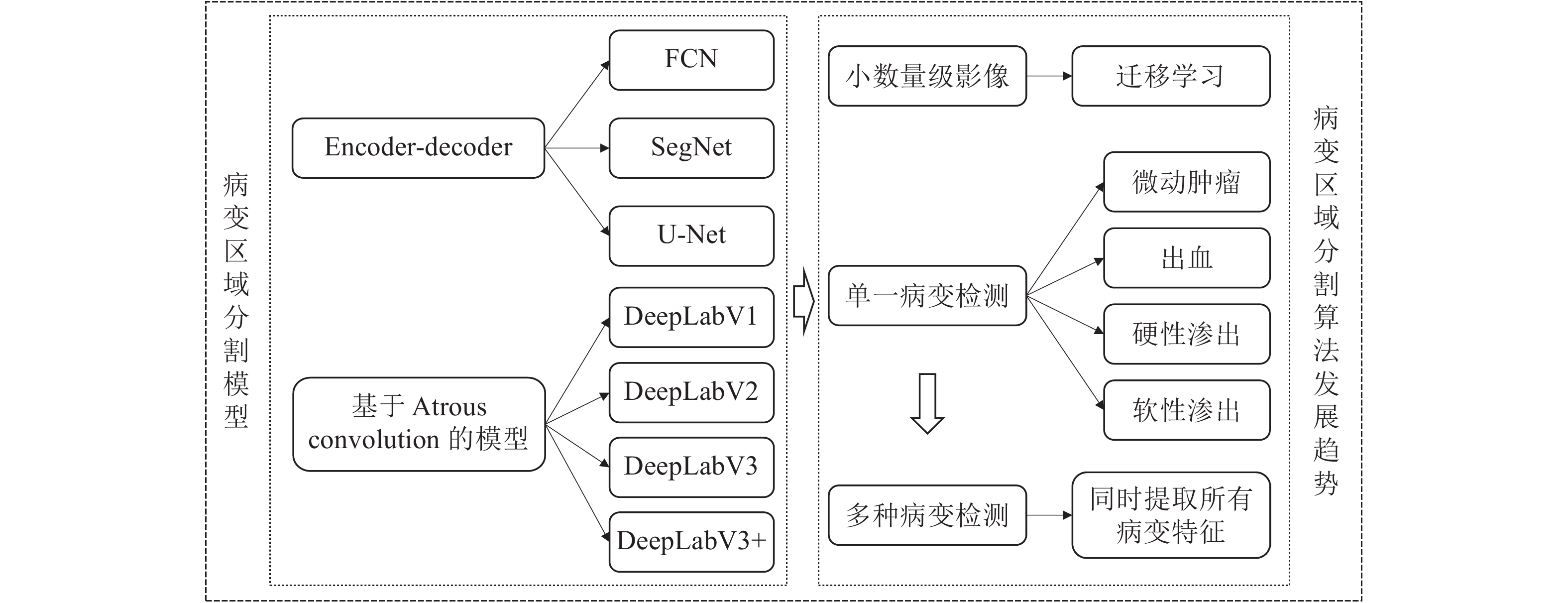
表 3 病变区域检测相关研究
Table 3 Related works on lesion detection
相关研究 方法 数据集 提取特征 性能 Shan 等[51] Patches + 堆叠稀疏自动编码(SSAE) + 迁移学习 DIARETDB MA 敏感性: 91.6%, F-score: 91.3%, 准确性: 91.38% Budak 等[55] 深度卷积神经网络 (DCNN) 在线挑战数据集 (ROC) MA 比赛分数为 0.221, 高于其他方法 Dai 等[54] Alex-Net 框架为基础的 MS-
CNN模型当地医院收集数据, DIARETDB1 MA 准确率: 96.1% Orlando 等[56] CNN + 手工工程特征 + 随机
森林 (Random forest) 分类器MESSIDOR E-Ophtha MA, HE AUC: CNN: 0.7912, 手工工程: 0.7325, CNN+手工工程: 0.8932 AUC: CNN: 0.8374, 手工工程: 0.8812, CNN+手工工程: 0.9031 van Grinsven 等[53] 动态选择抽样策略 (SeS,
NSeS) + 10 层 CNNKaggle MESSIDOR HE 敏感性: 84.8%, 特异性: 90.4%, AUC: 91.7%, 敏感性: 93.1%,
特异性: 91.5%, AUC: 97.9%Prentasic 等[57] DNN DRiDB EX, SE 敏感性: 78%, F-score: 78% Otálora 等[58] 基于LeNet网络 + EGL的主
动学习策略 + 迁移学习E-Ophtha EX, SE 敏感性: 99.8%, 特异性: 99.6%,
准确性: 99.6%Abbasi-Sureshjani 等[52] ResNet DIARETDB1, DR2, E-Ophtha EX, SE AUC 分别为: 96.5%, 97.2 %, 99.4% Badar 等[40] SegNet 模型为基础的 Auto-encoder 网络 Messidor MA, HE, EX 精确度: 99.24% (EX),
97.86% (HM), 88.65% (MA)ISBI 韩国 VRT 团队 DeepLab + U-Net DRiDB MA, HE, SE, EX F-score 分别为: 0.4951, 0.6804, 0.6995, 0.7127 ISBI 中国平安科技
Patech 团队DenseNet + DeepLab V3 DRiDB MA, HE, EX F-score 分别为: 0.474, 0.649, 0.885 ISBI 中国科大讯飞 U-Net + 注意力机制 + DeepLabV3 + DRiDB MA, HE, SE, EX F-score 分别为: 0.5017, 0.5588, 0.6588, 0.8741 Tan 等[59] DCNN CLEOPATRA MA, HM, HE, SE 敏感性: 87.58%, 特异性: 98.73%
下载: 导出CSV
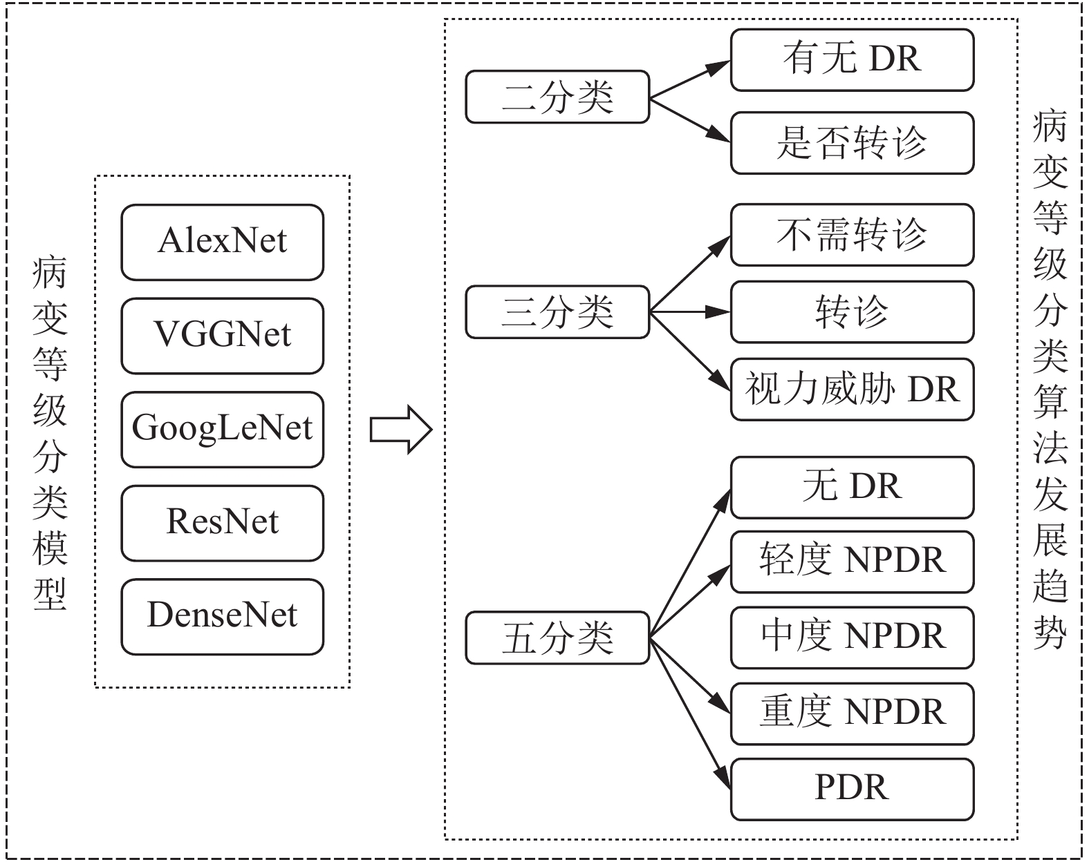
表 4 病变等级分类相关研究
Table 4 Related works on classification of diabetic retinopathy
相关研究 应用 方法 数据集 性能 谷歌 Gulshan 等[69] 诊断 RDR InceptionV3 框架,
端到端分类EyePACS-1
Messidor-2特异性: 93.4 %, 敏感性: 97.5 %
特异性: 93.9 %, 敏感性: 96.1%Li 等[72] 诊断有无 DR VggNet, GoogLeNet, Vgg-s 等进行迁移学习 DR1
MESSIDORVgg-s 性能最好:
敏感性: 97.11 %, 特异性: 86.03 %,
准确度: 92.01 %, AUC: 0.9834ElTanboly 等[73] 诊断 RDR Stacked non-negativityconstraint autoencoder (SNCAE) 医院获取 OCT 图像 准确度: 96 % Gargeya 等[74] 诊断 RDR Data-driven DNN ResNet, Second-level gradientboosting
classifierEyePACS
MESSIDORAUC: 0.97
AUC: 0.94Abràmoff 等[75] 诊断 RDR, VTDR DCNN Messidor-2 RDR 敏感性: 96.8 %, 特异性: 87.0 %,
AUC: 0.980
从 RDR 分类出 VTDR: 敏感性: 100 %,
特异性: 91 %, AUC: 0.989Ting 等[76] 诊断 RDR, VTDR VGG-19 6 个不同国家招募了 10
组数据集RDR 敏感性: 90.5 %, 特异性: 91.6 %,
AUC: 0.936
从 RDR 分类出 VTDR: 敏感性: 100 %,
特异性: 91.1 %, AUC: 0.958中山大学[16] 诊断 RDR, VTDR InceptionV3 中国人彩色眼底图片
多种族彩色眼底图像敏感性: 97.0 %, 特异性: 91.4 %
敏感性: 92.5 %, 特异性: 98.5 %Abràmoff 等[77] 在初级保健诊所诊断
DR, 进行实际应用CNN 在初级保健诊所招收了 900
名受试者, 男性占 47.5 %; 其
中包括: 西班牙裔 16.1 %, 非
裔美国人 28.6%敏感性: 87.2 %, 特异性: 90.7 %,
显像率: 96.1 %Wang等[78] 划分五类等级 DenseNet, Boosting
tree algorithmKaggle 精确度: R0: 0.92, R1: 0.70,
R2: 0.64, R3: 0.67, R4: 0.69Zhou 等[79] 划分五类等级 Multi-Cell Multi-Task CNN Kaggle Kappa: 0.841 Doshi 等[80] 划分五类等级 5层 CNN 网络 EyePACs Kappa: 0.386 IBM 划分五类等级 DCNN EyePAC 准确度: 86 %
下载: 导出CSV
表 5 基于视网膜OCT影像的眼部疾病诊断相关研究
Table 5 Studies on diagnosis of ocular diseases based on retinal OCT images
相关研究 应用 方法 数据集 性能 Sandhu 等[84] 对 DR 患者早期诊断 基于融合形状, 强度和空间信息的联合模型, 两阶段深度融合分类网络 路易斯维尔大学 (University of Louisville) 接受常规筛查和 (或) 监测检查的 II 型糖尿病患者 OCT 数据 准确率平均为94% Hassan等[81] 从 OCT 扫描中分割出
8 个视网膜层深度卷积神经网络, 基于结构张力的分割框架(CNN-STSF) 取自不同公共可用数据集和当地武装部队眼科研究所 (AFIO) 数据集的超过 3.9 万幅视网膜 OCT 影像 准确性: 93.75% Vahadane等[82] 分割硬渗出物和囊肿
区域检测 DME图像处理, 深度学习,
基于规则方法1827 幅 OCT 影像 Precision: 96.43%, Recall:
89.45%, F1-score: 92.81%Kermany 等[15] 疾病分类转诊 InceptionV3+ 迁移
学习, 分类网络108312 幅 OCT 训练影像 (37206幅脉络膜新生血管, 11349 幅糖尿病黄斑水肿,
8617 幅玻璃膜疣, 51140 幅正常),
1000 幅 OCT 测试影像 (每个类别 250 幅)准确率: 96.6 %, 敏感性: 97.8 %,
特异性: 97.4 %, AUC: 99.9 %Li 等[83] 疾病分类转诊 VGG-16 + 迁移学习,
分类网络医院获取 109312 幅 OCT 影像 (37456 幅脉络膜新生血管, 11599 幅糖尿病黄斑水肿, 8867 幅玻璃膜疣, 51390 幅正常) 准确率: 98.6%, 敏感性: 97.8%,
特异性: 99.4%, AUC: 100%DeepMind
团队[18]分割疾病特征
分类转诊3DU-NET 分割网络, CNN 分类网络 摩尔菲尔兹眼科医院提供 14 884 幅 OCT 影像 准确度: 94%, AUC: 99.21%
下载: 导出CSV
-
[1] Dhoot D S, Baker K, Saroj N, Vitti R, Berliner A J, Metzig C, et al. Baseline factors affecting changes in diabetic retinopathy severity scale score after intravitreal aflibercept or laser for diabetic macular edema: Post hoc analyses from VISTA and VIVID. Ophthalmology, 2018, 125(1): 51−56 doi: 10.1016/j.ophtha.2017.06.029 [2] 杨玲, 沈玺. 糖尿病性视网膜病变与干眼的相关性研究. 国际眼科杂志, 2018, 18(4): 744−747 doi: 10.3980/j.issn.1672-5123.2018.4.39Yang Ling, Shen Xi. Research on correlation between diabetic retinopathy and dry eye. International Eye Science, 2018, 18(4): 744−747 doi: 10.3980/j.issn.1672-5123.2018.4.39 [3] 张巧丽, 赵地, 迟学斌. 基于深度学习的医学影像诊断综述. 计算机科学, 2017, 44(S2): 1−7Zhang Qiao-Li, Zhao Di, Chi Xue-Bin. Review for deep learning based on medical imaging diagnosis. Computer Science, 2017, 44(S2): 1−7 [4] Ciulla T A, Amador A G, Zinman B. Diabetic retinopathy and diabetic macular edema: Pathophysiology, screening, and novel therapies. Diabetes Care, 2003, 26(9): 2653−2664 doi: 10.2337/diacare.26.9.2653 [5] 孙扬, 李琪欢, 田思佳, 张杰, 王友信, 平昭, 等. 免疫球蛋白G N-糖基化与糖尿病视网膜病变的相关性研究. 中国预防医学杂志, 2018, 19(10): 734−737Sun Yang, Li Qi-Huan, Tian Si-Jia, Zhang Jie, Wang You-Xin, Ping Zhao, et al. Association of immunoglobulin G N-glycosylation and diabetic retinopathy. China Preventive Medicine, 2018, 19(10): 734−737 [6] Palis A G, Golnik K C, Mayorga E P, Filipe H P, Garg P. The international council of ophthalmology 360-degree assessment tool: Development and validation. Canadian Journal of Ophthalmology, 2018, 53(2): 145−49 doi: 10.1016/j.jcjo.2017.09.002 [7] Kaur S, Singh D. Early detection and classification of diabetic retinopathy using empirical transform and SVM. In: Proceedings of the 2018 Computational Vision and Bio Inspired Computing. Coimbatore, India: Springer, 2018. 1072−1083 [8] Wong T Y, Sun J, Kawasaki R, Ruamviboonsuk P, Gupta N, Lansingh V C, et al. Guidelines on diabetic eye care: The international council of ophthalmology recommendations for screening, follow-up, referral, and treatment based on resource settings. Ophthalmology, 2018, 125(10): 1608−1622 doi: 10.1016/j.ophtha.2018.04.007 [9] Gass J D M, Sever R J, Sparks D, Goren J. A combined technique of fluorescein funduscopy and angiography of the eye. Archives of Ophthalmology, 1967, 78(4): 455−461 doi: 10.1001/archopht.1967.00980030457009 [10] Hernandez-Matas C, Zabulis X. Super resolution for fundoscopy based on 3D image registration. In: Proceedings of the 36th Annual International Conference of the IEEE Engineering in Medicine and Biology Society. Chicago, IL, USA: IEEE, 2014. 6332−6338 [11] 孙延奎. 光学相干层析医学图像处理及其应用. 光学精密工程, 2014, 22(4): 1086−1104 doi: 10.3788/OPE.20142204.1086Sun Yan-Kui. Medical image processing techniques based on optical coherence tomography and their applications. Optics and Precision Engineering, 2014, 22(4): 1086−1104 doi: 10.3788/OPE.20142204.1086 [12] Lin A D, Lee A Y, Zhang Q Q, Rezaei K A, Kinyoun J, Wang R K, et al. Association between OCT-based microangiography perfusion indices and diabetic retinopathy severity. British Journal of Ophthalmology, 2017, 101(7): 960−964 doi: 10.1136/bjophthalmol-2016-309514 [13] Eltanboly A H, Palacio A, Shalaby A M, Switala A E, Helmy O, Schaal S, et al. An automated approach for early detection of diabetic retinopathy using SD-OCT images. Frontiers in Bioscience, 2018, 10(1): 197−207 [14] Muiesan M L, Salvetti M, Paini A, Riviera M, Pintossi C, Bertacchini F, et al. Ocular fundus photography with a smartphone device in acute hypertension. Journal of Hypertension, 2017, 35(8): 1660−1665 doi: 10.1097/HJH.0000000000001354 [15] Kermany D S, Goldbaum M, Cai W J, Valentim C C S, Liang H Y, Baxter S L, et al. Identifying medical diagnoses and treatable diseases by image-based deep learning. Cell, 2018, 172(5): 1122−1131 doi: 10.1016/j.cell.2018.02.010 [16] Li Z X, Keel S, Liu C, He Y F, Meng W, Scheetz J, et al. An automated grading system for detection of vision-threatening referable diabetic retinopathy on the basis of color fundus photographs. Diabetes Care, 2018, 41(12): 2509−2516 doi: 10.2337/dc18-0147 [17] Mookiah M R K, Acharya U R, Chua C K, Lim C M, Ng E Y K, Laude A. Computer-aided diagnosis of diabetic retinopathy: A review. Computers in Biology & Medicine, 2013, 43(12): 2136−2155 [18] De Fauw J, Ledsam J R, Romera-Paredes B, Nikolov S, Tomasev N, Blackwell S, et al. Clinically applicable deep learning for diagnosis and referral in retinal disease. Nature Medicine, 2018, 24(9): 1342−1350 doi: 10.1038/s41591-018-0107-6 [19] IDX. Press release: FDA permits marketing of IDx-DR for automated detection of diabetic retinopathy in primary care [Online], available: https://www.eyediagnosis.net/press-releases/press-release-fda-permits-marketing-of-idx-dr-for-automated-detection-of-diabetic-retinopathy-in-primary-care, April 12, 2018 [20] Rajalakshmi R, Subashini R, Anjana R M, Mohan V. Automated diabetic retinopathy detection in smartphone-based fundus photography using artificial intelligence. Eye, 2018, 32(6): 1138−1144 doi: 10.1038/s41433-018-0064-9 [21] 郭潇雅. 嵩岳机器人惊艳亮相. 中国医院院长, 2018, (14): 28−29Guo Xiao-Ya. Songyue robot makes an amazing appearance. Chinese Hospital CEO, 2018, (14): 28−29 [22] Oloumi F, Rangayyan R M, Ells A L. Computer-aided diagnosis of proliferative diabetic retinopathy. In: Proceedings of the 2012 Annual International Conference of the IEEE Engineering in Medicine & Biology Society. San Diego, CA, USA: IEEE, 2012. 1438−1441 [23] Ting D S W, Cheung G C M, Wong T Y. Diabetic retinopathy: Global prevalence, major risk factors, screening practices and public health challenges: A review. Clinical & Experimental Ophthalmology, 2016, 44(4): 260−277 [24] Porwal P, Pachade S, Kamble R, Kokare M, Deshmukh G, Sahasrabuddhe V, et al. Indian diabetic retinopathy image dataset [Online], available: https://ieee-dataport.org/open-access/indian-diabetic-retinopathy-image-dataset-idrid, November 13, 2018 [25] Decenciére E. TeleOphta: Machine learning and image processing methods for teleophthalmology [Online], available: http://www.adcis.net/en/Download-Third-Party/E-Ophtha.html, January 10, 2013 [26] Prentașić P. Diabetic retinopathy image dataset [Online], available: https://ipg.fer.hr/ipg/resources/image_database, 2013. [27] French Ministry. Methods to evaluate segmentation and indexing techniques in the field of retinal ophthalmology [Online], available: http://www.adcis.net/en/Download-Third-Party/Messidor.html, 2014. [28] LaTIM laboratory. Messidor 2 [Online], available: http://latim.univ-brest.fr/indexfce0.html [29] Kauppi T, Kalesnykiene V, Kamarainen J K, Lensu L, Sorri I, Pietilä J, et al. DIARETDB0 — Standard diabetic retinopathy database: Calibration level 0 [Online], available: http://www.it.lut.fi/project/imageret/diaretdb0/index.html, May 30, 2007 [30] Kauppi T, Kalesnykiene V, Kamarainen J K, Lensu L, Sorri I, Raninen A, et al. DIARETDB1-Standard diabetic retinopathy database: Calibration level 1 [Online], available: http://www.it.lut.fi/project/imageret/diaretdb1/, June 19, 2007 [31] Rasti R, Rabbani H, Mehridehnavi A, Hajizadeh F. Macular OCT classification using a multi-scale convolutional neural network ensemble. IEEE Transactions on Medical Imaging, 2018, 37(4): 1024−1034 doi: 10.1109/TMI.2017.2780115 [32] 于观贞, 魏培莲, 陈颖, 朱明华. 人工智能在肿瘤病理诊断和评估中的应用与思考. 第二军医大学学报, 2017, 38(11): 1349−1354Yu Guan-Zhen, Wei Pei-Lian, Chen Ying, Zhu Ming-Hua. Artificial intelligence in pathological diagnosis and assessment of human solid tumor: Application and thinking. Academic Journal of Second Military Medical University, 2017, 38(11): 1349−1354 [33] Wu bo. AI medical imaging landing actual combat: Data annotation, algorithm method, computing power optimization [Online], available: http://www.mooc.ai/course/384/learn?lessonid=2165#lesson/2165, December 21, 2017 [34] Karger D R, Oh S, Shah D. Efficient crowdsourcing for multi-class labeling. ACM SIGMETRICS Performance Evaluation Review, 2013, 41(1): 81−92 doi: 10.1145/2494232.2465761 [35] Long J, Shelhamer E, Darrell T. Fully convolutional networks for semantic segmentation. In: Proceedings of the 2015 IEEE Conference on Computer Vision and Pattern Recognition. Boston, MA, USA: IEEE, 2015. 3431−3440 [36] Badrinarayanan V, Kendall A, Cipolla R. SegNet: A deep convolutional encoder-decoder architecture for image segmentation. IEEE Transactions on Pattern Analysis and Machine Intelligence, 2017, 39(12): 2481−2495 doi: 10.1109/TPAMI.2016.2644615 [37] Ronneberger O, Fischer P, Brox T. U-Net: Convolutional networks for biomedical image segmentation. In: Proceedings of the International Conference on Medical Image Computing and Computer-Assisted Intervention. Munich, Germany: Springer, 2015. 234−241 [38] Simonyan K, Zisserman A. Very deep convolutional networks for large-scale image recognition. arXiv preprint arXiv: 1409.1556, 2014. [39] He K M, Zhang X Y, Ren S Q, Sun J. Deep residual learning for image recognition. In: Proceedings of the 2016 IEEE Conference on Computer Vision and Pattern Recognition. Las Vegas, NV, USA: IEEE, 2016. 770−778 [40] Badar M, Shahzad M, Fraz M M. Simultaneous segmentation of multiple retinal pathologies using fully convolutional deep neural network. In: Proceedings of Annual Conference on Medical Image Understanding and Analysis. Southampton, United Kingdom: Springer, 2018. 313−324 [41] He K M, Zhang X Y, Ren S Q, Sun J. Spatial pyramid pooling in deep convolutional networks for visual recognition. In: Proceedings of European Conference on Computer Vision. Zurich, Switzerland: Springer, 2014. 346−361 [42] Chen L C, Papandreou G, Kokkinos I, Murphy K, Yuille A L. Semantic image segmentation with deep convolutional nets and fully connected CRFs. arXiv preprint arXiv: 1412.7062, 2014. [43] Chen L C, Papandreou G, Kokkinos I, Murphy K, Yuille A L. DeepLab: Semantic image segmentation with deep convolution al nets, atrous convolution, and fully connected CRFs. IEEE Transactions on Pattern Analysis and Machine Intelligence, 2018, 40(4): 834−848 [44] Lin T Y, Dollár P, Girshick R, He K M, Hariharan B, Belongie S. Feature pyramid networks for object detection. In: Proceed ings of the 2017 IEEE Conference on Computer Vision and Pat tern Recognition. Honolulu, HI, USA: IEEE, 2017. 936−944 [45] Chen L C, Papandreou G, Schroff F, Adam H. Rethinking atrous convolution for semantic image segmentation. arXiv preprint arXiv: 1706.05587, 2017. [46] Chen L C, Zhu Y K, Papandreou G, Schroff F, Adam H. Encoder-decoder with atrous separable convolution for semantic image segmentation. arXiv preprint arXiv: 1802.02611, 2018. [47] Chollet F. Xception: Deep learning with depthwise separable convolutions. In: Proceedings of the 2017 IEEE Conference on Computer Vision and Pattern Recognition. Honolulu, HI, USA: IEEE, 2017. 1800−1807 [48] Yosinski J, Clune J, Bengio Y, Lipson H. How transferable are features in deep neural networks? In: Proceedings of the 27th International Conference on Neural Information Processing Systems. Cambridge, MA, United States: MIT Press, 2014. 3320−3328 [49] Wang D X, Cui P, Zhu W W. Deep asymmetric transfer network for unbalanced domain adaptation. In: Proceedings of the 32nd AAAI Conference on Artificial Intelligence. New Orleans, Louisiana, USA: AAAI, 2018.443−450 [50] Dou Q, Ouyang C, Chen C, Chen H, Heng P A. Unsupervised cross-modality domain adaptation of convnets for biomedical image segmentations with adversarial loss. arXiv preprint arXiv: 1804.10916, 2018. [51] Shan J, Li L. A deep learning method for microaneurysm detec tion in fundus images. In: Proceedings of the 1st International Conference on Connected Health: Applications, Systems and En gineering Technologies (CHASE). Washington, DC: IEEE, 2016. 357−358 [52] Abbasi-Sureshjani S, Dashtbozorg B, ter Haar Romeny B M, Fleuret F. Boosted exudate segmentation in retinal images using residual nets. In: Proceedings of the 2017 Fetal, Infant and Ophthalmic Medical Image Analysis. Québec City, Canada: Springer, 2017. 210−218 [53] van Grinsven M J J P, van Ginneken B, Hoyng C B, Theelen T, Sánchez C I. Fast convolutional neural network training using selective data sampling: Application to hemorrhage detection in color fundus images. IEEE Transactions on Medical Imaging, 2016, 35(5): 1273−1284 [54] Dai L, Fang R G, Li H T, Hou X H, Sheng B, Wu Q, et al. Clinical report guided retinal microaneurysm detection with multi-sieving deep learning. IEEE Transactions on Medical Imaging, 2018, 37(5): 1149−1161 doi: 10.1109/TMI.2018.2794988 [55] Budak U, Şengür A, Guo Y H, Akbulut Y. A novel microaneurysms detection approach based on convolutional neural networks with reinforcement sample learning algorithm. Health Information Science and Systems, 2017, 5(1): 14 [56] Orlando J I, Prokofyeva E, del Fresno M, Blaschko M B. An ensemble deep learning based approach for red lesion detection in fundus images. Computer Methods and Programs in Biomedicine, 2018, 153: 115−127 [57] Prentasic P, Lonçarić S. Detection of exudates in fundus photographs using deep neural networks and anatomical landmark detection fusion. Computer Methods and Programs in Biomedicine, 2016, 137: 281−292 doi: 10.1016/j.cmpb.2016.09.018 [58] Otálora S, Perdomo O, González F, Müller H. Training deep convolutional neural networks with active learning for exudate classification in eye fundus images. In: Proceedings of the 2017 Intravascular Imaging and Computer Assisted Stenting, and Large-Scale Annotation of Biomedical Data and Expert Label Synthesis. Québec City, Canada: Springer, 2017. 146−154. [59] Tan J H, Fujita H, Sivaprasad S, Bhandary S V, Raod A K, Chua K C, et al. Automated segmentation of exudates, haemorrhages, microaneurysms using single convolutional neural network. Information Sciences, 2017, 420: 66−76 [60] Mansour R F. Evolutionary computing enriched computer-aided diagnosis system for diabetic retinopathy: A survey. IEEE Reviews in Biomedical Engineering, 2017, 10: 334−349 [61] LeCun Y, Bottou L, Bengio Y, Haffner P. Gradient-based learning applied to document recognition. Proceedings of the IEEE, 1998, 86(11): 2278−2324 [62] Vaswani A, Shazeer N, Parmar N, Uszkoreit J, Jones L, Gomez A N, et al. Attention is all you need. In: Proceedings of the 31st International Conference on Neural Information Processing Systems. Red Hook, NY, United States: Curran Associates Inc., 2017. 6000−6010 [63] Ioffe S, Szegedy C. Batch normalization: Accelerating deep network training by reducing internal covariate shift. In: Proceedings of the 2015 International Conference on Machine Learning. PMLR, 2015: 448−456 [64] Krizhevsky A, Sutskever I, Hinton G E. ImageNet classification with deep convolutional neural networks. In: Proceedings of the 25th International Conference on Neural Information Processing Systems. Red Hook, NY, United States: Curran Associates Inc., 2012. 1097−1105 [65] Szegedy C, Liu W, Jia Y Q, Sermanet P, Reed S, Anguelov D, et al. Going deeper with convolutions. In: Proceedings of the 2015 IEEE Conference on Computer Vision and Pattern Recognition. Boston, MA, USA: IEEE, 2015. 1−9 [66] Lu D H, Heisler M, Lee S, Ding G, Sarunic M V, Beg M F. Retinal fluid segmentation and detection in optical coherence tomography images using fully convolutional neural network. arXiv preprint arXiv: 1710.04778, 2017. [67] Ioffe S, Szegedy C. Batch normalization: Accelerating deep network training by reducing internal covariate shift. arXiv preprint arXiv: 1502.03167, 2015. [68] Szegedy C, Vanhoucke V, Ioffe S, Shlens J, Wojna Z. Rethinking the inception architecture for computer vision. In: Proceedings of the 2016 IEEE Conference on Computer Vision and Pattern Recognition. Las Vegas, USA: IEEE, 2016: 2818−2826 [69] Gulshan V, Peng L, Coram M, Stumpe M C, Wu D, Narayanaswamy A, et al. Development and validation of a deep learning algorithm for detection of diabetic retinopathy in retinal fundus photographs. JAMA, 2016, 316(22): 2402−2410 [70] Huang G, Liu Z, Van Der Maaten L, Weinberger K Q. Densely connected convolutional networks. In: Proceedings of the 2017 IEEE Conference on Computer Vision and Pattern Recognition. Honolulu, HI, USA: IEEE, 2017. 2261−2269 [71] Wong T Y, Bressler N M. Artificial intelligence with deep learning technology looks into diabetic retinopathy screening. JAMA, 2016, 316(22): 2366−2367 doi: 10.1001/jama.2016.17563 [72] Li X G, Pang T T, Xiong B, Liu W X, Liang P, Wang T F. Convolutional neural networks based transfer learning for diabetic retinopathy fundus image classification. In: Proceedings of the 10th International Congress on Image and Signal Processing, BioMedical Engineering and Informatics (CISP-BMEI). Shanghai, China: IEEE, 2017. 1−11 [73] ElTanboly A, Ghazaf M, Khalil A, Shalaby A, Mahmoud A, Switala A, et al. An integrated framework for automatic clinical assessment of diabetic retinopathy grade using spectral domain OCT images. In: Proceedings of the 15th International Symposium on Biomedical Imaging (ISBI 2018). Washington, DC, USA: IEEE, 2018. 1431−1435 [74] Gargeya R, Leng T. Automated identification of diabetic retinopathy using deep learning. Ophthalmology, 2017, 124(7): 962−969 doi: 10.1016/j.ophtha.2017.02.008 [75] Abràmoff M D, Lou Y Y, Erginay A, Clarida W, Amelon R, Folk J C, et al. Improved automated detection of diabetic retinopathy on a publicly available dataset through integration of deep learning. Investigative Ophthalmology & Visual Science, 2016, 57(13): 5200−5206 [76] Ting D S W, Cheung C Y L, Lim G, Tan G S W, Quang N D, Gan A, et al. Development and validation of a deep learning system for diabetic retinopathy and related eye diseases using retinal images from multiethnic populations with diabetes. JAMA, 2017, 318(22): 2211−2223 doi: 10.1001/jama.2017.18152 [77] Abràmoff M D, Lavin P T, Birch M, Shah N, Folk J C. Pivotal trial of an autonomous AI-based diagnostic system for detection of diabetic retinopathy in primary care offices. NPJ Digital Medicine, 2018, 1: 39 doi: 10.1038/s41746-018-0040-6 [78] Wang Y, Wang G A, Fan W G, Li J X. A deep learning based pipeline for image grading of diabetic retinopathy. In: Proceedings of the International Conference on Smart Health. Switzerland: Springer, 2018. 240−248 [79] Zhou K, Gu Z W, Liu W, Luo W X, Cheng J, Gao S H, et al. Multi-cell multi-task convolutional neural networks for diabetic retinopathy grading. In: Proceedings of the 40th Annual International Conference of the IEEE Engineering in Medicine and Biology Society (EMBC). Honolulu, HI, USA: IEEE, 2018. 2724−2727 [80] Doshi D, Shenoy A, Sidhpura D, Gharpure P. Diabetic retinopathy detection using deep convolutional neural networks. In: Proceedings of the 2016 International Conference on Computing, Analytics and Security Trends (CAST). Pune, India: IEEE, 2016. 261−266 [81] Hassan T, Usman A, Akram M U, Masood M F, Yasin U. Deep learning based automated extraction of intra-retinal layers for analyzing retinal abnormalities. In: Proceedings of the 20th International Conference on e-Health Networking, Applications and Services (Healthcom). Ostrava, Czech Republic: IEEE, 2018. 1−5 [82] Vahadane A, Joshi A, Madan K, Dastidar T R. Detection of diabetic macular edema in optical coherence tomography scans using patch based deep learning. In: Proceedings of the 15th International Symposium on Biomedical Imaging (ISBI 2018). Washington DC, USA: IEEE, 2018. 1427−1430 [83] Li F, Chen H, Liu Z, Zhang X D, Wu Z Z. Fully automated detection of retinal disorders by image-based deep learning. Graefe´s Archive for Clinical and Experimental Ophthalmology, 2019, 257(3): 495−505 [84] Sandhu H S, Eltanboly A, Shalaby A, Keynton R S, Schaal S, El-Baz A. Automated diagnosis and grading of diabetic retinopathy using optical coherence tomography. Investigative Ophthalmology & Visual Science, 2018, 59(7): 3155−3160 [85] Powers D M W. Evaluation: From precision, recall and F-measure to ROC, informedness, markedness and correlation. arXiv Preprint arXiv: 2010.16061, 2020. [86] Goutte C, Gaussier E. A probabilistic interpretation of precision, recall and F-score, with implication for evaluation. In: Proceedings of the European Conference on Information Retrieval. Santiago de Compostela, Spain: Springer, 2005. 345−359 [87] Zou K H, O´Malley A J, Mauri L. Receiver-operating characteristic analysis for evaluating diagnostic tests and predictive models. Circulation, 2007, 115(5): 654−657 doi: 10.1161/CIRCULATIONAHA.105.594929 [88] 吴江, 侯绍新, 靳萌萌, 胡忠义. 基于LDA模型特征选择的在线医疗社区文本分类及用户聚类研究. 情报学报, 2017, 36(11): 1183−1191 doi: 10.3772/j.issn.1000-0135.2017.11.010Wu Jiang, Hou Shao-Xin, Jin Meng-Meng, Hu Zhong-Yi. LDA feature selection based text classification and user clustering in Chinese online health community. Journal of the China Society for Scientific and Technical Information, 2017, 36(11): 1183−1191 doi: 10.3772/j.issn.1000-0135.2017.11.010 [89] Sim J, Wright C C. The kappa statistic in reliability studies: Use, interpretation, and sample size requirements. Physical Therapy, 2005, 85(3): 257−268 doi: 10.1093/ptj/85.3.257 [90] Graham B. Kaggle Diabetic Retinopathy Detection Competition Report, University of Warwick, UK, 2015. [91] 李曼华, 孙昊鹏, 尤启冬. CYP1A2抑制剂预测模型的建立及评价. 中国药科大学学报, 2013, 44(5): 401−409 doi: 10.11665/j.issn.1000-5048.20130504Li Man-Hua, Sun Hao-Peng, You Qi-Dong. Establishment and evaluation of prediction models for the discovery of CYP1A2 inhibitors. Journal of China Pharmaceutical University, 2013, 44(5): 401−409 doi: 10.11665/j.issn.1000-5048.20130504 [92] 杨蓓. 贝叶斯网络模型在老年人认知功能评价队列研究中的应用 [硕士学位论文], 山西医科大学, 中国, 2017.Yang Bei. Bayesian Network Modeling: A Cohort Study of Cognitive Function Evaluation among the Elderly [Master Thesis], Shanxi Medical University, China, 2017. [93] 田娟秀, 刘国才, 谷珊珊, 鞠忠建, 刘劲光, 顾冬冬. 医学图像分析深度学习方法研究与挑战. 自动化学报, 2018, 44(3): 401−424Tian Xiu-Juan, Liu Guo-Cai, Gu Shan-Shan, Ju Zhong-Jian, Liu Jin-Guang, Gu Dong-Dong. Deep learning in medical image analysis and its challenges. Acta Automatica Sinica, 2018, 44(3): 401−424 [94] Zheng T, Xie W, Xu L L, He X Y, Zhang Y, You M R, et al. A machine learning-based framework to identify type 2 diabetes through electronic health records. International Journal of Medical Informatics, 2017, 97: 120−127 doi: 10.1016/j.ijmedinf.2016.09.014 [95] 郑光远, 刘峡壁, 韩光辉. 医学影像计算机辅助检测与诊断系统综述. 软件学报, 2018, 29(5): 1471−1514Zheng Guang-Yuan, Liu Xia-Bi, Han Guang-Hui. Survey on medical image computer aided detection and diagnosis systems. Journal of Software, 2018, 29(5): 1471−1514 [96] Rahu M, McKee M. Epidemiological research labelled as a violation of privacy: The case of Estonia. International Journal of Epidemiology, 2008, 37(3): 678−682 doi: 10.1093/ije/dyn022 [97] Tramèr F, Zhang F, Juels A, Reiter M K, Ristenpart T. Stealing machine learning models via prediction APIs. In: Proceedings of the 25th USENIX Conference on Security Symposium. Berkeley, CA, United States: USENIX Association, 2016. 601−618 [98] Shokri R, Stronati M, Song C Z, Shmatikov V. Membership inference attacks against machine learning models. In: Proceedings of the 2017 IEEE Symposium on Security and Privacy (SP). San Jose, CA, USA: IEEE, 2017. 3−18 [99] Abadi M, Chu A, Goodfellow I, McMahan H B, Mironov I, Talwar K, et al. Deep learning with differential privacy. In: Proceedings of the 2016 ACM SIGSAC Conference on Computer and Communications Security. Vienna, Austria: ACM, 2016. 308−318 [100] Doshi-Velez F, Kim B. Towards a rigorous science of interpretable machine learning. arXiv preprint arXiv:1702.08608, 2017. [101] Goodman B, Flaxman S. European union regulations on algorithmic decision-making and a right to explanation. arXiv Preprint arXiv: 1606.08813, 2016. [102] Lum K, Isaac W. To predict and serve? Significance, 2016, 13(5): 14−19 doi: 10.1111/j.1740-9713.2016.00960.x [103] Faber J, Fonseca L M. How sample size influences research outcomes. Dental Press Journal of Orthodontics, 2014, 19(4): 27−29 doi: 10.1590/2176-9451.19.4.027-029.ebo -

 下载:
下载:

计量
- 文章访问数: 3276
- HTML全文浏览量: 2574
- PDF下载量: 735
- 被引次数: 0
